Review
. 2021 Feb;73(1):53-63.
doi: 10.1007/s00251-020-01196-0. Epub 2021 Jan 11.

Anatomy of teleost fish immune structures and organs

Affiliations
Review

Anatomy of teleost fish immune structures and organs

Håvard Bjørgen et al. Immunogenetics. 2021 Feb.

Abstract

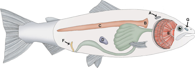
The function of a tissue is determined by its construction and cellular composition. The action of different genes can thus only be understood properly when seen in the context of the environment in which they are expressed and function. We now experience a renaissance in morphological research in fish, not only because, surprisingly enough, large structures have remained un-described until recently, but also because improved methods for studying morphological characteristics in combination with expression analysis are at hand. In this review, we address anatomical features of teleost immune tissues. There are approximately 30,000 known teleost fish species and only a minor portion of them have been studied. We aim our review at the Atlantic salmon (Salmo salar) and other salmonids, but when applicable, we also present information from other species. Our focus is the anatomy of the kidney, thymus, spleen, the interbranchial lymphoid tissue (ILT), the newly discovered salmonid cloacal bursa and the naso-pharynx associated lymphoid tissue (NALT).

Keywords: Bursa; Fish; Histology; ILT; Immune organ; Kidney; Lymphoid tissue; Morphology; Spleen; Thymus.


Figures

Fig. 1
Fig. 1
Schematic topography of immune organs in Atlantic salmon. A Thymus, B head kidney, C trunk kidney, D spleen, E gills with the interbranchial lymphoid tissue (ILT), F salmonid bursa, G olfactory organ with the nasopharynx-associated lymphoid tissue (NALT)
Fig. 2
Fig. 2
Micrograph of thymus, transverse sections, Atlantic salmon, HE stain. a Overview with gill chamber (GC) and underlying musculature (MU). Note the well-developed pharyngeal epithelium (PE) and the capsule (C). b Higher magnification shows pharyngeal epithelium (PE), outer zone (OZ), inner zone (IZ), and subcapsular zone (SZ). Arrow indicates a longitudinal section of a septa which might be mistaken for a Hassall’s corpuscle in a transverse section
Fig. 3
Fig. 3
Kidney, Atlantic salmon, HE stain. a The head kidney is dominated by hematopoietic interstitial tissue. Note the abundance of melano-macrophages (black). Endocrine tissue (adrenal homolog) is marked with asterisk. b The transition between head kidney (HK) and trunk kidney (TK) is marked with a red, dotted line. Note the appearance of glomeruli and tubuli in the trunk kidney. c The trunk kidney is dominated by exocrine tissue with nephrons and tubuli. Multiple glomeruli are present in the image (arrows). The interstitium contains hematopoietic tissue and melano-macrophages (black)
Fig. 4
Fig. 4
Spleen, Atlantic salmon, HE stain. a At low magnification, the histological difference between the white pulp (W) and the red pulp (R) is more prominent than at higher magnification. b Higher magnification allows identification of melano-macrophages (arrow, black cells) in the white pulp (W) whereas no such cells are present in the red pulp (R). c At high magnification of the white pulp (W), an ellipsoid containing an erythrocyte (arrowhead) can be observed together with scattered melano-macrophages (arrow, black cells)
Fig. 5
Fig. 5
Interbranchial lymphoid tissue (ILT) of Atlantic salmon. a Micrograph showing that the ILT is divided into a proximal (pILT) and distal (dILT) compartments consisting of reticular epithelial cells with pockets containing predominantly T lymphocytes. b Transmission electron microscopy (TEM) image of the epithelial surface of the pILT showing flattened epithelial cells with microridges (arrows) partly covering a goblet cell (asterisk). c TEM image showing the basal membrane (arrows) and epithelial cells (EC) of the pILT in a sexually mature Atlantic salmon. Note the protrusions of the basal membrane interacting with the proximal cell membranes of the epithelial cells of the stratum basale and the interdigitating lateral cellular extensions of these cells. This construction enhances the attachment of the pILT to the basal membrane and solidifies the structure. The construction does not seem to facilitate transport over the basal membrane
Fig. 6
Fig. 6
The salmonid bursa, Atlantic salmon. a Macroscopic location of the bursa caudal to the urogenital papilla (large arrow). Note the anal labiae (small arrows) which may be closed in vivo. b Macroscopic observation of the salmonid bursa, sagittal section. Bursa (arrow), the orifices of the urogenital papilla (arrowhead) and hindgut (HG) are all marked. c The bursa (arrow) may be opened with forceps for a closer inspection. Note the whitish epithelium which may be indicative for a high content of leukocytes. The urogenital papilla (arrowhead) and hind gut (HG) are marked. d Micrograph, transverse section, HE stained. The bursa terminates in two sacks with prominent lymphoepithelium (arrows). The ducts of the urogenital papilla (U/G) and the hindgut (HG) are seen in the section. e Micrograph, transverse section, HE stained, mid part of the salmonid bursa. Note the prominent lymphoepithelium (LE). f Micrograph, transverse section, cytokeratin immunostain, mid part of the salmonid bursa. Note the epithelial meshwork (red) in which lymphocytes are embedded
Fig. 7
Fig. 7
Sections from the olfactory organ, Atlantic salmon, HE stained. a A primary lamella (PL) with secondary lamellae (SL and boxed). Note that the primary lamella is filled with nervous tissue and abundant melanin-containing cells (arrowhead). b Enlarged image of a secondary lamella showing the two epithelial compartments; tips (arrowhead) and olfactory epithelium (arrows). c In the olfactory epithelium, lymphocytes may be observed (white arrowhead). Note the cilia on the epithelial cells (black arrowheads)

References

    1. Aas IB, et al. Transcriptional characterization of the T cell population within the salmonid interbranchial lymphoid tissue. J Immunol. 2014;193(7):3463–3469. doi: 10.4049/jimmunol.1400797. - DOI - PubMed
    1. Aas IB, et al. The interbranchial lymphoid tissue likely contributes to immune tolerance and defense in the gills of Atlantic salmon. Dev Comp Immunol. 2017;76:247–254. doi: 10.1016/j.dci.2017.06.013. - DOI - PubMed
    1. Abelli L et al (1998) Apoptosis of thymocytes in developing sea bass Dicentrarchus labrax (L.). Fish Shellfish Immunol 8(1):13–24
    1. Agius C, Roberts R. Melano-macrophage centres and their role in fish pathology. J Fish Dis. 2003;26(9):499–509. doi: 10.1046/j.1365-2761.2003.00485.x. - DOI - PubMed
    1. Austbø L, et al. Transcriptional response of immune genes in gills and the interbranchial lymphoid tissue of Atlantic salmon challenged with infectious salmon anaemia virus. Dev Comp Immunol. 2014;45(1):107–114. doi: 10.1016/j.dci.2014.02.007. - DOI - PubMed

MeSH terms

LinkOut - more resources