Review
. 2021 Feb 10;121(3):1845-1893.
doi: 10.1021/acs.chemrev.0c00981. Epub 2021 Jan 11.

Proteostasis of Islet Amyloid Polypeptide: A Molecular Perspective of Risk Factors and Protective Strategies for Type II Diabetes

Affiliations
Review

Proteostasis of Islet Amyloid Polypeptide: A Molecular Perspective of Risk Factors and Protective Strategies for Type II Diabetes

Danilo Milardi et al. Chem Rev. .

Abstract

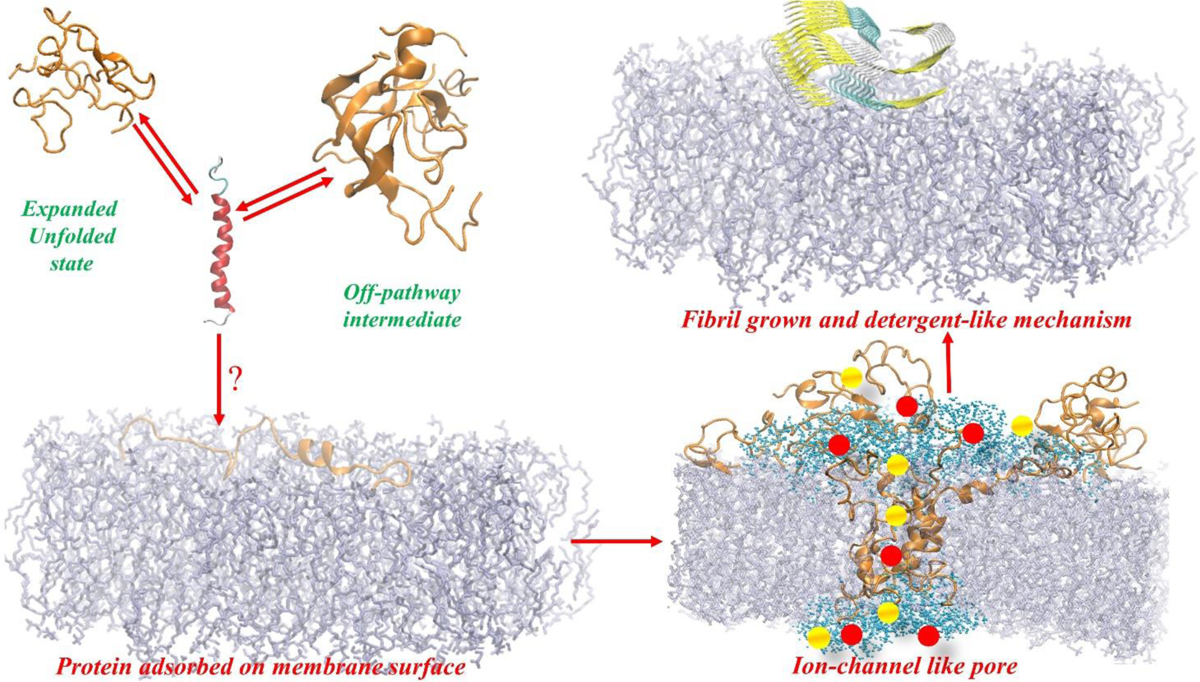
The possible link between hIAPP accumulation and β-cell death in diabetic patients has inspired numerous studies focusing on amyloid structures and aggregation pathways of this hormone. Recent studies have reported on the importance of early oligomeric intermediates, the many roles of their interactions with lipid membrane, pH, insulin, and zinc on the mechanism of aggregation of hIAPP. The challenges posed by the transient nature of amyloid oligomers, their structural heterogeneity, and the complex nature of their interaction with lipid membranes have resulted in the development of a wide range of biophysical and chemical approaches to characterize the aggregation process. While the cellular processes and factors activating hIAPP-mediated cytotoxicity are still not clear, it has recently been suggested that its impaired turnover and cellular processing by proteasome and autophagy may contribute significantly toward toxic hIAPP accumulation and, eventually, β-cell death. Therefore, studies focusing on the restoration of hIAPP proteostasis may represent a promising arena for the design of effective therapies. In this review we discuss the current knowledge of the structures and pathology associated with hIAPP self-assembly and point out the opportunities for therapy that a detailed biochemical, biophysical, and cellular understanding of its aggregation may unveil.


Figures

Figure 1.
Figure 1.
A) Human islet from a diabetic subject where most of the β-cells are replaced by amyloid. Congo red- Bar 50 μm. B) β-cell with intracellular amyloid-containing secretory granules. White arrows point to amyloid. C) Proposed sequence of events leading to islet amyloidosis: a) First, the processing of proIAPP is affected by factors such as high levels of NEFAs or glucose. Granules of amyloid-like fibrils fuse and form proIAPP amyloid deposits; b) Over time this aggregate enlarges and replaces most of the cell; c) the affected cell dies and the amyloid becomes extracellular and can act as a template for further amyloid formation; d) amyloid is now made up by IAPP secreted from neighboring β-cells. Formation of extracellular amyloid is preceded by the formation of cytotoxic intermediates, which can interact with cell membrane of surrounding cells and cause ion influx triggering apoptosis. This Figure is reproduced with copyright permission from Reference 130.
Figure 2.
Figure 2.. Amino acid sequence of IAPP from various species.
The amino acid sequence of IAPP is highly conserved with a disulfide bond between Cys-2 and Cys-7 residues and an amidated C-terminus. The peptide region spanning residues 20 to 29 shows the highest sequence variation among the species which is highlighted in the box.
Figure 3.
Figure 3.. Studies of sequence alterations on the aggregation of full-length hIAPP and truncated IAPP8–37.
Amino acid substitutions in red show higher aggregation propensity as compared to wild-type hIAPP, with a shorter lag-time; substitutions in blue show a lower aggregation propensity with an extended lag-time or no aggregation; substitutions in green did not change the aggregation behavior as compared to that of the wild-type hIAPP. X is 2-aminobutyric acid (2-Abu). Cam is carboxyamidomethyl protecting group. N-MeG and N-MeI are N-methylation of the peptide bonds at Gly-24 and Ile-26. isoD is isoAsp.
Figure 4.
Figure 4.
Schematic diagram illustrating a RAGE-mediated mechanism of hIAPP cytotoxicity in β-cells.
Figure 5.
Figure 5.
Schematic illustration of a two-step membrane disruption mechanism. In the aqueous phase, the oligomers, formed on-pathway and off-pathway, are in chemical equilibrium with the monomers existing in α-helix conformation. In the first step hIAPP insert as α-helix within the lipid bilayer, then the peptides self-assemble to form a pentameric aggregate having an ion-channel like structure which can enable ions to cross the bilayer resulting in an electrolytic imbalance. In the second step, monomers from the solution can associate with the lipid bilayer surface which can catalyze the self-assembly process to form fibrils; the fibril forming process can remove lipids from one of the leaflets of the bilayer and then ripping the other leaflet causing membrane damage like a “detergent-like” carpet mechanism.
Figure 6.
Figure 6.
Schematic illustration of lipid-assisted bilayer penetration. Chemical equilibrium between dispersed lipid monomers (or free lipids) in solution and their supramolecular assemblies (LUVs) is always established and is characterized by the Critical Micellar Concentration (CMC). For lipid molecules having short hydrocarbon tails or charged head-groups, the concentration of free lipids in equilibrium with LUVs may reach values up to the μM range, while the CMC of long-tails containing lipids the CMC value drops to few nM. In the aqueous phase, three simultaneous and competing equilibria should be taken into account: lipid-lipid, protein-protein and lipid-protein with the monomeric species. Lipids having a high CMC value favour the formation of a lipid-protein complex. The lipid-protein complex transfers into the lipid bilayer spontaneously due to the low chemical potential of the lipid bilayer when compared to that of the aqueous phase. Thus, lipids with a high CMC value would favour ion-channel like pores formation whereas lipids having a low CMC value would favour a detergent-like mechanism and fibril formation.
Figure 7.
Figure 7.
Human-IAPP degradation by proteases. Solid lines represent the cleavage sites targeted by the proteases. The yellow highlighted residues 22–27 form the amyloidogenic core of hIAPP.
Figure 8.
Figure 8.
Chemical structures of the multifunctional compounds discussed in the text.

References

    1. Schwartz P New patho-anatomic observations on amyloidosis in the aged. Fluorescence microscopic investigations. In Amyloidosis; Mandema E, Ruinen L, Scholten JH, Cohen AS, Eds.; Excerpta Medica: Amsterdam, 1968; pp 400–15.
    1. Westermark P Islet Amyloid Polypeptide and Amyloid in the Islets of Langerhans; Leslie RDG, Robbins D, Eds.; Cambridge University Press: Cambridge, UK, 1995.
    1. Sengupta U; Nilson AN; Kayed R The Role of Amyloid-β Oligomers in Toxicity, Propagation, and Immunotherapy. EBioMedicine 2016, 6, 42–49. - PMC - PubMed
    1. Scollo F; La Rosa C Amyloidogenic Intrinsically Disordered Proteins: New Insights into Their Self-Assembly and Their Interaction with Membranes. Life 2020, 10, 144. - PMC - PubMed
    1. Klaips CL; Jayaraj GG; Hartl FU Pathways of Cellular Proteostasis in Aging and Disease. J. Cell Biol 2018, 217, 51–63. - PMC - PubMed

Publication types

Substances