Review
. 2021 Mar;78(6):2429-2457.
doi: 10.1007/s00018-020-03706-5. Epub 2021 Jan 11.

Cerebrospinal fluid outflow: a review of the historical and contemporary evidence for arachnoid villi, perineural routes, and dural lymphatics

Affiliations
Review

Cerebrospinal fluid outflow: a review of the historical and contemporary evidence for arachnoid villi, perineural routes, and dural lymphatics

Steven T Proulx. Cell Mol Life Sci. 2021 Mar.

Abstract

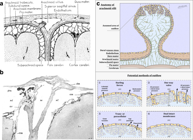
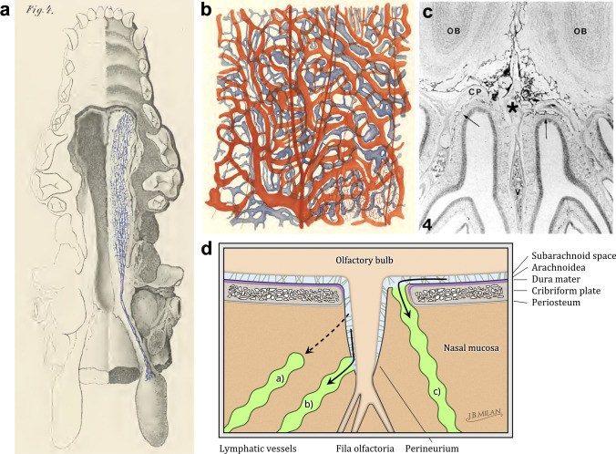
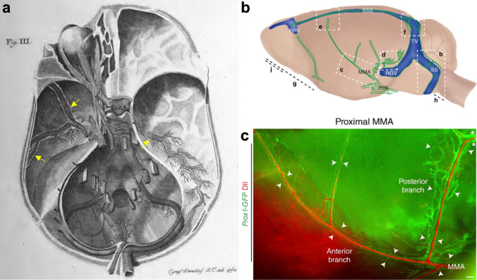
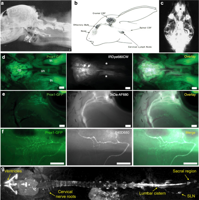
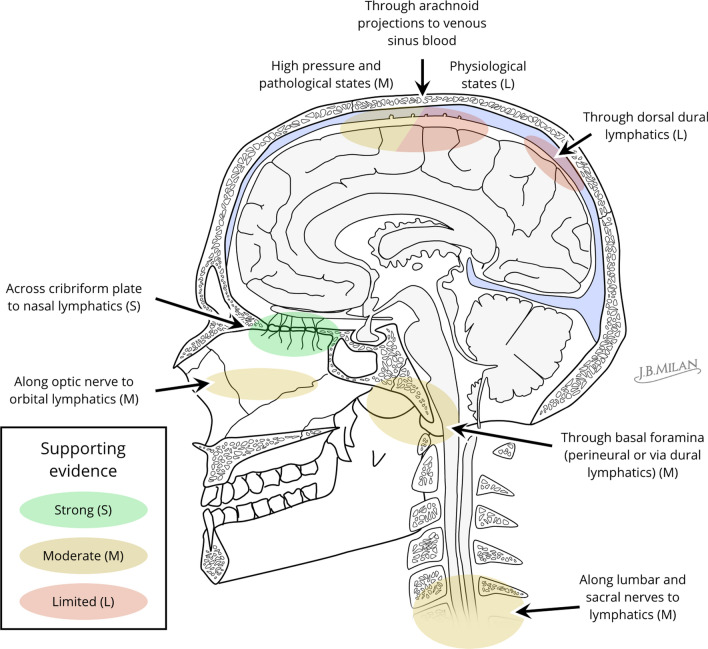
Cerebrospinal fluid (CSF) is produced by the choroid plexuses within the ventricles of the brain and circulates through the subarachnoid space of the skull and spinal column to provide buoyancy to and maintain fluid homeostasis of the brain and spinal cord. The question of how CSF drains from the subarachnoid space has long puzzled scientists and clinicians. For many decades, it was believed that arachnoid villi or granulations, outcroppings of arachnoid tissue that project into the dural venous sinuses, served as the major outflow route. However, this concept has been increasingly challenged in recent years, as physiological and imaging evidence from several species has accumulated showing that tracers injected into the CSF can instead be found within lymphatic vessels draining from the cranium and spine. With the recent high-profile rediscovery of meningeal lymphatic vessels located in the dura mater, another debate has emerged regarding the exact anatomical pathway(s) for CSF to reach the lymphatic system, with one side favoring direct efflux to the dural lymphatic vessels within the skull and spinal column and another side advocating for pathways along exiting cranial and spinal nerves. In this review, a summary of the historical and contemporary evidence for the different outflow pathways will be presented, allowing the reader to gain further perspective on the recent advances in the field. An improved understanding of this fundamental physiological process may lead to novel therapeutic approaches for a wide range of neurological conditions, including hydrocephalus, neurodegeneration and multiple sclerosis.

Keywords: CSF; Clearance; Cranial nerves; Cribriform plate; Lymphatic vessels; Meningeal.


Conflict of interest statement

The author declares that he has no conflict of interest.

Figures

Fig. 1
Fig. 1
Arachnoid projections and the different models that have been proposed for CSF outflow to the dural venous sinus. a Lewis Weed’s rendering of an arachnoid villus (from [263]). The interior of the villus is composed of fluid spaces continuous with the subarachnoid space and contains fibers that that are connected with the arachnoid trabeculae. The villus projects through dural tissue to extend into the lumen of the superior sagittal sinus. A cap of arachnoid cells covered by an intact layer of endothelial cells is located at the terminus of the villus. b A reproduction of an image from Lewis Weed’s published report from 1914 [3] showing Prussian blue precipitate (gm, black granular material) within an arachnoid villus (av) extending toward the superior sagittal sinus (ss). Granules are seen in “isolated arachnoidal clumps near the sinus lumen”. The image is acquired from a cat that had been injected with 2% potassium ferrocyanide and iron ammonium citrate into the lumbar space under a pressure of 180 mm H2O for four hours. Note the large subdural space (sd) between the arachnoid mater (am) and dura mater (dm) which is an artifact of the tissue preparation. Weed also highlights the “absence of precipitate within the true dura tissue surrounding the arachnoidal elements” representing early evidence for the arachnoid barrier layer. c Various models attempting to provide a mechanism for outflow of CSF and solutes through arachnoid projections (villi or granulations) have been proposed. Panel 1 is Weed’s original conception based upon Starlings forces in which favorable hydrostatic and osmotic gradients would exist through the endothelial cell lining to facilitate CSF outflow to the dural venous sinus (DVS). Hugh Davson later challenged this concept as no osmotic forces would develop to allow macromolecular solutes, such as albumin, to leave the CSF [52]. Panel 2 is the proposal of arachnoid granulations (AG) as one-way valves, as conceived by Welch and Friedman [60]. When hydrostatic pressure gradients are favorable, endothelial-lined channels at the end of the projection are open for flow of fluid and macromolecules to the venous sinus. When the pressure in the sinus is above that of the SAS, then the channels are collapsed and no flow in either direction can occur. These endothelial-lined channels were later identified using electron microscopy as infoldings of the tissue that were not continuous with the SAS. Panel 3 are the various models that were proposed to explain how flow might occur through an intact layer of endothelial cells on the cap of the projections. Panel 4 represents the concept of a dual layer of intact cells on the cap of the projections, one layer comprised of arachnoid cells and the other of venous endothelium. In this scenario, no outflow of CSF would normally occur through these structures under homeostatic conditions. However, these layers may be distended and eventually disrupted under conditions of high intracranial pressure, allowing for a “safety valve” mechanism for rapid egress of CSF. Scheme by Joachim Birch Milan
Fig. 2
Fig. 2
CSF outflow through the cribriform plate to nasal lymphatics. a Reproduction of an image from Key and Retzius [2] showing the lower surface of a dog's skull in which the lower maxilla and palate have been removed on one side to show the lymphatic vessels of the nasal and palatal submucosa filled with Richardson's blue dye from the SAS. The dye is transported to the deep cervical lymph glands of the neck. b Also from Key and Retzius [2], demonstrating Richardson's blue dye-filled lymphatics in the nasal submucosa of a rabbit injected into the “subdural” space of the brain. The authors noted that similar filling of vessels was observed in animals after injections into the SAS. c Reproduction of an image from Kida et al. [116] showing a decalcified coronal section from a rat that had been injected with India ink. Connections through the cribriform plate are evident with India ink particles found within lymphatic vessels on the nasal submucosa side of the plate (arrows). d Models for anatomical routes of CSF outflow to reach lymphatic vessels in the nasal tissue. Experimental support exists for three anatomical routes for tracers to cross the cribriform plate alongside the olfactory nerves. Model (a) represents an outflow pathway along a contiguous subarachnoid–perineural space (light blue). In this model, the perineurium is a continuation of the arachnoid mater, however, it does not form a barrier outside of the skull or is only loosely adhered to the nerve. Fluid and solutes have unrestricted access to the interstitium of the nasal (sub)mucosa (brown) where they then enter the lymphatic vasculature within this tissue (yellow). In model (b), CSF also crosses the plate through a perineural space but then has a direct pathway to lymphatic capillaries that surround the nerve in a collar-like fashion. Model (c) shows bona fide lymphatic vessels crossing the cribriform plate to directly access the CSF on the CNS side. In this model the arachnoid mater would not form a barrier in the region covering the cribriform plate. Scheme by Joachim Birch Milan
Fig. 3
Fig. 3
Dural lymphatic vessels of the basal skull in mice and humans. a Reproduction of a plate from Mascagni, 1787 [165]. Using injections of colored beeswax and mercury in human cadavers, dural lymphatic vessels (lighter colored vessels indicated by yellow arrows) were identified at the base of the skull alongside the middle meningeal arteries and veins and were found to leave the skull with these vessels at the foramina spinosa. b Schematic from Aspelund et al. [144] demonstrating regions of the dura found to contain lymphatic vessels in the Prox1-GFP transgenic reporter mouse. MMA middle meningeal artery, PPA pterygopalatine artery, RGV retroglenoid vein, RRV rostral rhinal vein, SS sigmoid sinus, SSS superior sagittal sinus, TV transverse vein. c Close up of the region c shown in panel b, indicating the proximity of the Prox1-GFP lymphatic vessels (green) to the branches of middle meningeal arterial (MMA) vessel network [perfused via intravenous DiI dye injection (red)] in an analagous situation to that described by Mascagni in humans
Fig. 4
Fig. 4
In vivo imaging techniques for assessment of outflow of CSF tracers. a Reproduction from Mortensen and Sullivan, 1933 [90]. The image shows a radiograph of a dog 6 h after injection into the cisterna magna with thorotrast. The contrast agent is seen within the lymphatic vessels and the superficial and deep lymph nodes. b An image reprinted from Pile-Spellman et al. [110] showing a scintigraphic image of a rabbit that was injected into the lateral ventricle with 99mTc antimony sulfide. The sagittal image was acquired 4 h after injection and shows clear enrichment of radiotracer within the nasal region as well as in the deep cervical lymph node. c Inferior posterior radiograph of a cat skull 30 min after infusion of Isovist 300 contrast medium into the cisterna magna. Besides an accumulation in the nasal region (N), contrast medium (asterisk) is apparent in the orbital tissue and around the length of the optic nerve (O). Reproduced from [122] d–f Fluorescence microscopy imaging of the outflow of near-infrared fluorescent tracers from the CNS in Prox1-GFP transgenic reporter mice after intraventricular injection. Panel d indicates accumulation of IRDye680CW dye at the cribriform plate (cp, white arrows) and around the optic nerves (on, white asterisks). tn trigeminal nerves. In e, 3 kDa dextran labeled with AlexaFluor680 is shown exiting the orbit of the eye within Prox1-GFP+ lymphatic vessels. From this location the tracer drains into the mandibular (aka superficial cervical) lymph nodes (not shown). Panel f demonstrates an outflow of a 40 kDa pegylated near-infrared dye (P40D680) from the sacral region of the spine into Prox1-GFP+ lymphatic vessels. g Imaging with T1-weighted MRI of Gadospin D contrast agent showing spread to and outflow from the spine after low-rate infusion (0.1 μl/min) into the lateral ventricle. The outflow from the caudal end of the spine to the sacral lymph nodes is apparent. Panels d and e are reproduced from [130], while f and g are taken from [184]
Fig. 5
Fig. 5
An overview of the evidence for routes of CSF outflow. A qualitative assessment of the level of supporting evidence for various CSF outflow pathways was made based on the number of published studies, uniformity of the results across different species and plausibility of the anatomical and physiological mechanisms. Experimental support appears to be strongest for an outflow of CSF at the cribriform plate with lymphatic vessels carrying the fluid and solutes to the deep cervical lymph nodes. Outflow also appears to be evident at the optic nerves and at the base of the skull through several possible foramina either in a perineural manner or through dural lymphatics. Evidence also exists for outflow from the spine to lymphatic vessels, particularly in the lumbar or sacral regions. CSF outflow has been detected in a limited number of studies in mice to the dural lymphatic vessels on the superior aspect of the skull. There is little support for an outflow to arachnoid villi or granulations under normal conditions, however, this route may become recruited during conditions of increased intracranial pressure. Scheme by Joachim Birch Milan

References

    1. Engelhardt B, Vajkoczy P, Weller RO. The movers and shapers in immune privilege of the CNS. Nat Immunol. 2017;18(2):123–131. doi: 10.1038/ni.3666. - DOI - PubMed
    1. Key A, Retzius G. Studien in der Anatomie des Nervensystems und des Bindegewebes. Stockholm: Samson & Waller; 1875.
    1. Weed LH. Studies on cerebro-spinal fluid. No. III : the pathways of escape from the subarachnoid spaces with particular reference to the arachnoid villi. J Med Res. 1914;31(1):51–91. - PMC - PubMed
    1. Weed LH. Studies on cerebro-spinal fluid. No. IV: the dual source of cerebro-spinal fluid. J Med Res. 1914;31(1):93–118. - PMC - PubMed
    1. Davson H, Segal MB. Physiology of the CSF and blood–brain barriers. Boca Raton: CRC Press; 1996.