Review
. 2021 Mar;15(1):25-56.
doi: 10.1007/s12079-020-00602-2. Epub 2021 Jan 11.

CCN2 (Cellular Communication Network factor 2) in the bone marrow microenvironment, normal and malignant hematopoiesis

Affiliations
Review

CCN2 (Cellular Communication Network factor 2) in the bone marrow microenvironment, normal and malignant hematopoiesis

Roos J Leguit et al. J Cell Commun Signal. 2021 Mar.

Abstract

CCN2, formerly termed Connective Tissue Growth Factor, is a protein belonging to the Cellular Communication Network (CCN)-family of secreted extracellular matrix-associated proteins. As a matricellular protein it is mainly considered to be active as a modifier of signaling activity of several different signaling pathways and as an orchestrator of their cross-talk. Furthermore, CCN2 and its fragments have been implicated in the regulation of a multitude of biological processes, including cell proliferation, differentiation, adhesion, migration, cell survival, apoptosis and the production of extracellular matrix products, as well as in more complex processes such as embryonic development, angiogenesis, chondrogenesis, osteogenesis, fibrosis, mechanotransduction and inflammation. Its function is complex and context dependent, depending on cell type, state of differentiation and microenvironmental context. CCN2 plays a role in many diseases, especially those associated with fibrosis, but has also been implicated in many different forms of cancer. In the bone marrow (BM), CCN2 is highly expressed in mesenchymal stem/stromal cells (MSCs). CCN2 is important for MSC function, supporting its proliferation, migration and differentiation. In addition, stromal CCN2 supports the maintenance and longtime survival of hematopoietic stem cells, and in the presence of interleukin 7, stimulates the differentiation of pro-B lymphocytes into pre-B lymphocytes. Overexpression of CCN2 is seen in the majority of B-acute lymphoblastic leukemias, especially in certain cytogenetic subgroups associated with poor outcome. In acute myeloid leukemia, CCN2 expression is increased in MSCs, which has been associated with leukemic engraftment in vivo. In this review, the complex function of CCN2 in the BM microenvironment and in normal as well as malignant hematopoiesis is discussed. In addition, an overview is given of data on the remaining CCN family members regarding normal and malignant hematopoiesis, having many similarities and some differences in their function.

Keywords: Bone marrow; CCN2; CTGF; Connective tissue growth factor; Hematopoiesis; Leukemogenesis.


Conflict of interest statement

The authors report no conflict of interest.

Figures

Fig. 1
Fig. 1
CCN2 protein structure. Schematic representation of the full length CCN2 protein, which is made up by a signal peptide and 4 protein domains, depicted by the blue cylinders. Domain 1 consists of the insulin-like growth factor binding protein (IGFBP) module and domain 2 contains the von Willebrand factor C (VWC) module, together forming the N-terminal fragment of the protein. Domain 3 consists of the thrombospondin type 1 repeat (TSP1) module and domain 4 contains the C-terminal cysteine knot (CT) module, together forming the C-terminal fragment of the protein. The N- and C-terminal fragments are joint by a hinge region. Between the different domains, multiple cleavage sites are present, were CCN2 is cleaved by proteases, plasmin, chymotrypsin and matrix metalloproteinases. Loss of the signal peptide leads to intracellular retention of the protein. The protein contains 2 glycosylation sites. The functional relevance of glycosylation is, however, still unknown. Other (not depicted) posttranscriptional and posttranslational modifications to which the protein is subject to, are splicing, regulation by miRNAs and multimerisation
Fig. 2
Fig. 2
Overview of the complex regulation of CCN2 expression, and the diverse actions of the CCN2 protein, as reported in literature. Regulation of CCN2 expression. a Factors inducing CCN2 expression include growth factors, coagulation factors, hormones, bioactive lipids, glucose metabolism related factors, hypoxia and mechanical stress. b Factors inhibiting CCN2 expression include increased levels of cyclic adenosine monophosphate (cAMP), cytokines, insulin‐like growth factor binding protein‐4 (IGFBP-4), CCN3 and hepatic growth factor. c CCN2 expression is modified by posttranscriptional and posttranslational factors, which includes splicing, regulation by miRNAs, glycosylation, proteolytic cleavage and multimerisation of the protein. d CCN2 can induce its own expression by auto-induction, resulting in a positive feedback loop. Actions of the CCN2 protein: CCN2 exerts its function by binding to growth factors and cell surface receptors, thereby affecting intracellular signaling, as well as by binding to extracellular matrix (ECM) and other proteins. e CCN2 binds to growth-differentiation factor 5 (GDF-5), platelet derived growth factor (PDGF), insulin-like growth factor-1 and 2 (IGF1/2) and transforming growth factor-β (TGF-β). The effect of CCN2 on the binding of these ligands to their putative receptors is not fully elucidated. f CCN2 binds to bone morphogenetic proteins (BMPs), vascular endothelial growth factors (VEGFs) and fibroblast growth factor 2 (FGF2), thereby modulating their presentation and binding to cell-surface receptors, resulting in inhibited signal transduction. *Only full-length CCN2 inhibits VEGFA and VEGFC, not its proteolytic fragments. g CCN2 binds directly to structural ECM proteins, matricellular proteins and other proteins, including aggrecan, fibronectin, decorin, perlecan, CCN3, heparin, Wnt inhibitory factor 1 (Wif-1), Slit guidance ligand 3 (Slit-3), and von Willebrand factor (vWF). h CCN2 can bind directly to cell surface receptors, which include heparan sulphate proteoglycans (HSPGs), tropomyosin receptor kinase A (TrkA), dendritic cell-specific transmembrane protein (DC-STAMP), integrins, insulin-like growth factor 2 receptor (IGF-2R), epidermal growth factor receptor (EGFR), formyl peptide receptor-like 1 (FPRL1), osteoprotegerin (OPG), lipoprotein receptor-related proteins (LRPs), receptor activator of NF-κB (RANK), and fibroblast growth factor receptors (FGFRs). By CCN2 binding, intracellular signaling pathways and their cross-talk can be altered and gene transcription affected. i Intracellular signaling pathways that are affected by CCN2 include the extracellular signal–regulated kinase 1 and 2 (ERK1/2) pathway, the Rho GTPase pathway, the WNT pathway, the c-Jun N-terminal kinase (JNK) pathway, the phosphatidylinositol 3‑kinase (PI3K)/AKT pathway and the nuclear factor κB (NFκB) pathway the signaling mother against dexapentaplegic peptides (Smad) pathway. j CCN2 has been reported to act intracellularly after being taken up into the cytosol via endocytic pathways and, after phosphorylation, into the nucleus where it may affect gene transcription
Fig. 3
Fig. 3
CCN2 in the bone marrow microenvironment. CCN2 mRNA, depicted by formula image , is present in different bone marrow (BM) mesenchymal cells, including endothelial cells, osteoblasts, adipocytes and fibroblasts, with highest levels (formula image 
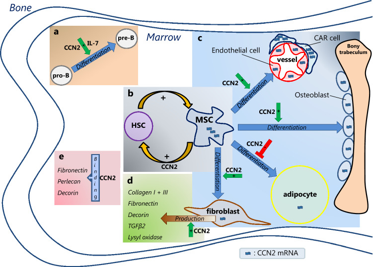
) reported in mesenchymal stem/stromal cell (MSC) and CXCL12-abundant reticular (CAR) cells. CCN2 exerts different actions in the BM. a In the presence of interleukin 7 (IL-7), CCN2 promotes pro-B cell to pre-B cell differentiation. b CCN2 produced by MSCs affects the (long-term) qualities of hematopoietic stem cells (HSCs). HSCs, in turn, upregulate CCN2 expression by MSCs. c CCN2 enhances the differentiation of MSCs into endothelial cells, osteoblast and fibroblasts, but has an inhibitory effect on the differentiation of MSCs into adipocytes. d CCN2 might induce the production of the ECM proteins collagen type I and type III, fibronectin, decorin, TGFβ-2 and lysyl oxidase by fibroblast. e CCN2 binds to fibronectin, perlecan and decorin, known constituents of the BM extracellular matrix. The effects hereof in the BM are yet unknown.
Figure 4
Figure 4
a Direct and indirect intracellular effects of CCN proteins in chronic myeloid leukemia (CML). CCN1 expression is increased in CML cells, inhibiting apoptosis through enhanced expression of the anti-apoptotic protein BCL2 via the NF-κB pathway, without involvement of AKT or ERK1/2. CCN3 expression is decreased in CML cells, decreasing its inhibitory effect of CCN3 on ERK and AKT phosphorylation, resulting in elevated levels of phosphorylated ERK and AKT. This leads to less apoptosis, presumably via the NF-κB pathway. In addition, decreased CCN3 levels result in less caspase 3 cleavage, thereby also reducing apoptosis. Furthermore, decreased CCN3 levels lead to less inhibition of NOTCH1 signaling, resulting in higher levels of NOTCH. This results in decreased expression of p27, disrupting cell cycle regulation. b Direct and indirect intracellular effects of CCN proteins in acute lymphoblastic leukemia (ALL). The effects have been demonstrated for CCN1 in B- and T-ALL, for CCN2 in B-ALL and for CCN4 in T-ALL cells. In these cells, CCN1, CCN2 as well as CCN4 expression is increased, all inhibiting apoptosis through enhanced expression of the anti-apoptotic protein BCL2. Both CCN1 and CCN2 activate AKT, whereas CCN4 activates AKT as well as ERK1/2. Involvement of the NF-κB pathway has been demonstrated for CCN1 and CCN4. In addition, increased CCN2 and CCN4 levels lead to decreased expression of the pro-apoptotic proteins BIM and BAX, respectively, both resulting in less apoptosis. Furthermore, increased CCN2 levels are associated with decreased p27 expression, thereby affecting cell cycle regulation

References

    1. Abd El Kader T, Kubota S, Nishida T, Hattori T, Aoyama E, Janune D, Hara ES, Ono M, Tabata Y, Kuboki T, Takigawa M. The regenerative effects of CCN2 independent modules on chondrocytes in vitro and osteoarthritis models in vivo. Bone. 2014;59:180–188. doi: 10.1016/j.bone.2013.11.010. - DOI - PubMed
    1. Abou-Shady M, Friess H, Zimmermann A, Di Mola FF, Guo XZ, Baer HU, Büchler MW. Connective tissue growth factor in human liver cirrhosis. Liver. 2000;20:296–304. doi: 10.1034/j.1600-0676.2000.020004296.x. - DOI - PubMed
    1. Abraham DJ, Shiwen X, Black CM, Sa S, Xu Y, Leask A. Tumor necrosis factor alpha suppresses the induction of connective tissue growth factor by transforming growth factor-beta in normal and scleroderma fibroblasts. J Biol Chem. 2000;275:15220–15225. doi: 10.1074/jbc.275.20.15220. - DOI - PubMed
    1. Abreu JG, Ketpura NI, Reversade B, De Robertis EM. Connective-tissue growth factor (CTGF) modulates cell signalling by BMP and TGF-beta. Nat Cell Biol. 2002;4:599–604. doi: 10.1038/ncb826. - DOI - PMC - PubMed
    1. Acar M, Kocherlakota KS, Murphy MM, Peyer JG, Oguro H, Inra CN, Jaiyeola C, Zhao Z, Luby-Phelps K, Morrison SJ. Deep imaging of bone marrow shows non-dividing stem cells are mainly perisinusoidal. Nature. 2015;526:126–130. doi: 10.1038/nature15250. - DOI - PMC - PubMed