. 2021 Aug;16(5):1297-1305.
doi: 10.1007/s11739-020-02596-6. Epub 2021 Jan 11.

A simple lung ultrasound protocol for the screening of COVID-19 pneumonia in the emergency department

Affiliations

A simple lung ultrasound protocol for the screening of COVID-19 pneumonia in the emergency department

Alessandro Dacrema et al. Intern Emerg Med. 2021 Aug.

Abstract

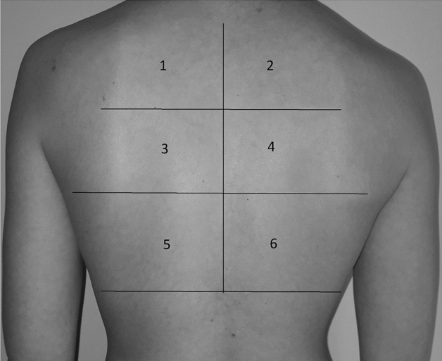
The most relevant manifestation of coronavirus disease 2019 (COVID-19) is interstitial pneumonia. Several lung ultrasound (US) protocols for pneumonia diagnosis are used in clinical practice, but none has been proposed for COVID-19 patients' screening in the emergency department. We adopted a simplified 6-scan lung US protocol for COVID-19 pneumonia diagnosis (LUSCOP) and compared its sensitivity with high resolution computed tomography (HRCT) in patients suspected for COVID-19, presenting to one Emergency Department from February 21st to March 15th, 2020, during the outbreak burst in northern Italy. Patients were retrospectively enrolled if both LUSCOP protocol and HRCT were performed in the Emergency Department. The sensitivity of LUSCOP protocol and HRCT were compared. COVID-19 pneumonia's final diagnosis was based on real-time reverse-transcription polymerase chain reaction from nasal-pharyngeal swab and on clinical data. Out of 150 suspected COVID-19 patients, 131 were included in the study, and 130 had a final diagnosis of COVID-19 pneumonia. The most frequent lung ultrasonographic features were: bilateral B-pattern in 101 patients (77%), B-pattern with subpleural consolidations in 26 (19.8%) and lung consolidations in 2 (1.5%). LUSCOP Protocol was consistent with HRCT in correctly screening 130 out of the 131 COVID-19 pneumonia cases (99.2%). In one case COVID-19 pneumonia was excluded by both HRCT and lung US. LUSCOP protocol showed optimal sensitivity and can be proposed as a simple screening tool for COVID-19 pneumonia diagnosis in the context of outbreak burst areas where prompt isolation of suspected patients is crucial for patients' and operators' safety.

Keywords: COVID-19; Emergency; Lung ultrasound; Outbreak; Pneumonia; Screening.


Conflict of interest statement

The authors declare that they have no conflict of interest.

Figures

Fig. 1
Fig. 1
Chest fields for lung US examination. Lung US scan protocol of posterior fields from the apex to the bases bilaterally: 6 fields in total, 3 fields for hemithorax, apex, middle, basal field.
Fig. 2
Fig. 2
Flowchart of patients’ enrollment. Hundred-fifty consecutive patients suspected for COVID-19 were recruited. For 6 patients, nasal-pharyngeal swab was lost and 6 patients had a negative result, therefore 12 patients had not COVID-19 confirmed diagnosis and were excluded. Of the 138 remaining patients, 129 patients had positive rRT-PCR from nasal-pharyngeal swab, and 9 had clinical diagnosis of COVID-19. Of the 129 patients, 5 patients had a false negative result from the first swab and a subsequent diagnostic confirmation from the second swab. Therefore, 138 patients had COVID-19 diagnostic confirmation. Of the 138 patients, 4 were excluded because HRCT was not performed and 3 because lung US was either not performed or not recorded on the medical file. Finally, 19 patients were excluded and 131 patients with confirmed COVID-19 diagnosis that underwent both lung US and HRCT were enrolled.
Fig. 3
Fig. 3
Comparison between HRCT and lung US according to LUSCOP protocol. From top to bottom: upper, medium, and lower fields are shown both in HRCT slices and in the corresponding right and left lung US. HRCT shows diffuse pulmonary emphysema mostly in upper and medium fields, associated with COVID-19 bilateral dorsal, subpleural ground-glass opacities, and consolidations. In the upper fields, B-lines at the lung US correspond to areas of mild interstitial involvement. In the middle fields, lung ultrasound shows confluent B-lines with pleural thickening. In the lower fields, pleural effusion is detected by lung US in the right side, associated with pleural thickening and irregularity; the left lung US shows a predominantly A-pattern corresponding to relatively spared parenchyma.
Fig. 4
Fig. 4
Comparison between HRCT and lung US according to LUSCOP protocol. From top to bottom: upper, medium, and lower fields are shown both in HRCT slices and in the corresponding right and left lung US. In this case, HRCT shows extensive COVID-19 pneumonia involving both lungs, with subpleural involvement, partially sparing the anterior and upper fields. Specifically, crazy paving pattern superimposition on ground glass opacities had an incremental cranio-caudal distribution with higher density in the subpleural dorsal fields. In the upper fields, lung US shows spared parenchyma. In the medium and lower fields, lung US shows an increasing B-pattern severity with confluent B lines and progressive pleural thickening with pre-consolidative state.

References

    1. https://www.who.int/emergencies/diseases/novel-coronavirus-2019/events-a.... Accessed April 20, 2020.
    1. Guan WJ, Ni ZY, Hu Y, et al. Clinical characteristics of coronavirus disease 2019 in China. N Engl J Med. 2020 doi: 10.1056/NEJMoa2002032. - DOI - PMC - PubMed
    1. Coronavirus disease (COVID-19) situation reports. Situation report 25. 14 February 2020. http://who.int/emergencies/diseases/novel-coronavirus-2019/situation-rep.... Accessed April 20, 2020.
    1. Chinese clinical guidance for COVID-19 pneumonia diagnosis and treatment (7th edition). March 2020. http://kjfy.meetingchina.org/msite/news/show/cn/3337.html. Accessed April 20, 2020.
    1. Fang Y, Zhang H, Xie J, Lin M, Ying L, Pang P, Ji W. Sensitivity of chest CT for COVID-19: comparison to RT-PCR. Radiology. 2020;19:200432. - PMC - PubMed

MeSH terms

LinkOut - more resources