

Knockdown of CENPF inhibits the progression of lung adenocarcinoma mediated by ERβ2/5 pathway
- PMID: 33428600
- PMCID: PMC7880349
- DOI: 10.18632/aging.202303
Knockdown of CENPF inhibits the progression of lung adenocarcinoma mediated by ERβ2/5 pathway
Abstract
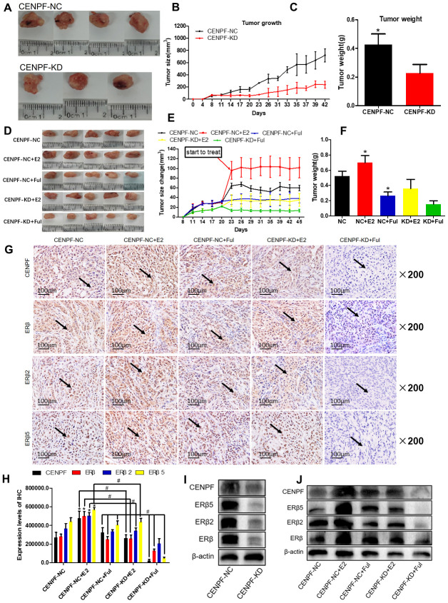
Many studies have reported that estrogen (E2) promotes lung cancer by binding to nuclear estrogen receptors (ER), and altering ER related nuclear protein expressions. With the GEO database analysis, Human centromere protein F (CENPF) is highly expressed in lung adenocarcinoma (LUAD), and the co-expression of CENPF and ERβ was found in the nucleus of LUAD cells through immunofluorescence. We identified the nuclear protein CENPF and explored its relationship with the ER pathway. CENPF and ERβ2/5 were related with T stage and poor prognosis (P<0.05). CENPF knockout significantly inhibited LUAD cell growth, the tumor growth of mice and the expression of ERβ2/5 (P<0.05). The protein expression of CENPF and ERβ2/5 in the CENPF-Knockdown+Fulvestrant group was lower than CENPF- Negative Control +Fulvestrant group (P=0.002, 0.004, 0.001) in A549 cells. The tumor size and weight of the CENPF-Knockdown+Fulvestrant group were significantly lower than CENPF- Negative Control +Fulvestrant group (P=0.001, 0.039) in nude mice. All the results indicated that both CENPF and ERβ2/5 play important roles in the progression of LUAD, and knockdown CENPF can inhibit the progression of LUAD by inhibiting the expression of ER2/5. Thus, the development of inhibitors against ERβ2/5 and CENPF remained more effective in improving the therapeutic effect of LUAD.
Keywords: WGCNA package; centromere protein F (CENPF); estrogen receptor beta; lung adenocarcinoma (LUAD); non-small cell lung cancer (NSCLC).
Conflict of interest statement
Figures


Similar articles
-
Overexpression of CENPF is associated with progression and poor prognosis of lung adenocarcinoma.Int J Med Sci. 2021 Jan 1;18(2):494-504. doi: 10.7150/ijms.49041. eCollection 2021. Int J Med Sci. 2021. PMID: 33390818 Free PMC article.
-
17β-estradiol upregulates IL6 expression through the ERβ pathway to promote lung adenocarcinoma progression.J Exp Clin Cancer Res. 2018 Jul 3;37(1):133. doi: 10.1186/s13046-018-0804-5. J Exp Clin Cancer Res. 2018. PMID: 29970138 Free PMC article.
-
Actin-like protein 8 promotes cell proliferation, colony-formation, proangiogenesis, migration and invasion in lung adenocarcinoma cells.Thorac Cancer. 2020 Mar;11(3):526-536. doi: 10.1111/1759-7714.13247. Epub 2020 Jan 21. Thorac Cancer. 2020. PMID: 31962007 Free PMC article.
-
Screening and identification of biomarkers associated with the diagnosis and prognosis of lung adenocarcinoma.J Clin Lab Anal. 2020 Oct;34(10):e23450. doi: 10.1002/jcla.23450. Epub 2020 Jul 16. J Clin Lab Anal. 2020. PMID: 32672359 Free PMC article.
-
LPCAT1 promotes brain metastasis of lung adenocarcinoma by up-regulating PI3K/AKT/MYC pathway.J Exp Clin Cancer Res. 2019 Feb 21;38(1):95. doi: 10.1186/s13046-019-1092-4. J Exp Clin Cancer Res. 2019. PMID: 30791942 Free PMC article.
Cited by
-
Combined multiomics analysis reveals the mechanism of CENPF overexpression-mediated immune dysfunction in diffuse large B-cell lymphoma in vitro.Front Genet. 2022 Dec 30;13:1072689. doi: 10.3389/fgene.2022.1072689. eCollection 2022. Front Genet. 2022. PMID: 36644760 Free PMC article.
-
Long Non-Coding RNA Small Nucleolar RNA Host Gene 4 Induced by Transcription Factor SP1 Promoted the Progression of Nasopharyngeal Carcinoma Through Modulating microRNA-510-5p/Centromere Protein F Axis.Biochem Genet. 2023 Oct;61(5):1967-1986. doi: 10.1007/s10528-023-10351-7. Epub 2023 Mar 10. Biochem Genet. 2023. PMID: 36899270
-
A Novel Prognostic Signature for Survival Prediction and Immune Implication Based on SARS-CoV-2-Related Genes in Kidney Renal Clear Cell Carcinoma.Front Bioeng Biotechnol. 2022 Jan 24;9:744659. doi: 10.3389/fbioe.2021.744659. eCollection 2021. Front Bioeng Biotechnol. 2022. PMID: 35141213 Free PMC article.
-
Lung Cancer Gene Regulatory Network of Transcription Factors Related to the Hallmarks of Cancer.Curr Issues Mol Biol. 2023 Jan 5;45(1):434-464. doi: 10.3390/cimb45010029. Curr Issues Mol Biol. 2023. PMID: 36661515 Free PMC article.
-
N6-methyladenosine modification of CENPF mRNA facilitates gastric cancer metastasis via regulating FAK nuclear export.Cancer Commun (Lond). 2023 Jun;43(6):685-705. doi: 10.1002/cac2.12443. Epub 2023 May 31. Cancer Commun (Lond). 2023. PMID: 37256823 Free PMC article.
References
-
- Chang GC, Tseng CH, Hsu KH, Yu CJ, Yang CT, Chen KC, Yang TY, Tseng JS, Liu CY, Liao WY, Hsia TC, Tu CY, Lin MC, et al.. Predictive factors for EGFR-tyrosine kinase inhibitor retreatment in patients with EGFR-mutated non-small-cell lung cancer - a multicenter retrospective SEQUENCE study. Lung Cancer. 2017; 104:58–64. 10.1016/j.lungcan.2016.12.002 - DOI - PubMed
-
- Anraku M, Waddell TK, de Perrot M, Lewis SJ, Pierre AF, Darling GE, Johnston MR, Zener RE, Rampersaud YR, Shepherd FA, Leighl N, Bezjak A, Sun AY, et al.. Induction chemoradiotherapy facilitates radical resection of T4 non-small cell lung cancer invading the spine. J Thorac Cardiovasc Surg. 2009; 137:441–47.e1. 10.1016/j.jtcvs.2008.09.035 - DOI - PubMed
-
- Ciuleanu TE, Ahmed S, Kim JH, Mezger J, Park K, Thomas M, Chen J, Poondru S, VanTornout JM, Whitcomb D, Blackhall F. Randomised phase 2 study of maintenance linsitinib (OSI-906) in combination with erlotinib compared with placebo plus erlotinib after platinum-based chemotherapy in patients with advanced non-small cell lung cancer. Br J Cancer. 2017; 117:757–66. 10.1038/bjc.2017.226 - DOI - PMC - PubMed
Publication types
MeSH terms
Substances
LinkOut - more resources
Full Text Sources
Other Literature Sources
Medical
