Review
. 2021 Sep:84:100940.
doi: 10.1016/j.preteyeres.2021.100940. Epub 2021 Jan 8.

The innate immune system in diabetic retinopathy

Affiliations
Review

The innate immune system in diabetic retinopathy

Warren W Pan et al. Prog Retin Eye Res. 2021 Sep.

Abstract

The prevalence of diabetes has been rising steadily in the past half-century, along with the burden of its associated complications, including diabetic retinopathy (DR). DR is currently the most common cause of vision loss in working-age adults in the United States. Historically, DR has been diagnosed and classified clinically based on what is visible by fundoscopy; that is vasculature alterations. However, recent technological advances have confirmed pathology of the neuroretina prior to any detectable vascular changes. These, coupled with molecular studies, and the positive impact of anti-inflammatory therapeutics in DR patients have highlighted the central involvement of the innate immune system. Reminiscent of the systemic impact of diabetes, immune dysregulation has become increasingly identified as a key element of the pathophysiology of DR by interfering with normal homeostatic systems. This review uses the growing body of literature across various model systems to demonstrate the clear involvement of all three pillars of the immune system: immune-competent cells, mediators, and the complement system. It also demonstrates how the relative contribution of each of these requires more extensive analysis, including in human tissues over the continuum of disease progression. Finally, although this review demonstrates how the complex interactions of the immune system pose many more questions than answers, the intimately connected nature of the three pillars of the immune system may also point to possible new targets to reverse or even halt reverse retinopathy.

Keywords: Complement system; Diabetic retinopathy; Innate immunity; Microglia and macroglia; Neurodegeneration; Neurovascular unit.


Conflict of interest statement

The authors have declared that no conflict of interest exists.

Figures

Figure 1.
Figure 1.. Anatomical blood-retinal barrier.
The layers of the retina, retinal pigment epithelium (RPE) and choroid are labeled on the left with details of the outer blood retinal barrier (oBRB) expanded in the diagram on the right depicting the tight and adherent junctions between the RPE cells in red. Additionally, a cartoon of the inner BRB (iBRB) is shown in the lower right with tight junction between endothelial cells, which are surrounded by pericytes, Müller glia, and astrocytes. Additionally, neurons of the ONL (outer nuclear layer) and INL (inner nuclear layer) are nearby. GCL: ganglion cell layer.
Figure 2.
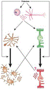
Figure 2.. The complex multi-cellular dysfunction in diabetic retinopathy.
Diabetes virtually affects all retinal cells in a sequence that remains hard to elucidate as they function as a neurovascular unit and influence each other. (1) Vessel dysfunction reflects pericytes and endothelial cells dysfunction and loss, a phenomenon influenced by factors secreted by dying neurons and activated micro- and macroglia. (2) Microglia (brown) and macroglia (green) become activated in response to the alteration of the retinal environment associated with diabetes, a phenomenon enhanced by neuronal cell death and vascular preturbations. (3) Alteration of the retinal homeostasis by diabetes leads to neuronal dysfunction and ultimately cell death, a phenomenon enhanced by the increasingly pro-inflammatory environment and vascular perturbations.
Figure 3.
Figure 3.. Microglial activation during progression of diabetic retinopathy.
The quiescent microglia observed in normal physiology is ramified in form and initially responds by becoming activated and transforming into an amoeboid state. During this appropriate response to diabetes, activated microglia secrete increasing amounts of neurotrophic and anti-inflammatory mediators and help preserve the state of no diabetic retinopathy. Over time and in absence of resolution of the unbalanced environment, the population of M2 appropriately active microglia shifts to a more M1 disease amplifying state, which participates in the progression of DR.
Figure 4.
Figure 4.. Macroglial activation during progression of diabetic retinopathy.
Quiescent macroglia become activated in response to diabetes and while first involved in the adaptive protective response, will ultimately contribute to diabetic retinopathy pathology. The exact nature and duration of this intermediary state between quiescent macroglia and activated macroglia remains to be fully elucidated but may be a key element in our quest to new avenues for DR treatment.
Figure 5.
Figure 5.. AlphaA-crystallin chaperone protein while increasingly expressed is decreasingly phosphorylated on a key regulatory site in DR donors.
AlphaA-crystallin total protein level was assessed by western-blot and showed to be increased in diabetic donors with or without DR (left). Concomitantly, phosphorylation on T148, a phosphosite controlling its protective function was assessed by multiple reaction monitoring and shown to be dramatically reduced in DR donors. (Adapted from Ruebsam et al., 2018)
Figure 6.
Figure 6.. IDO expression is elevated in the retina of diabetic donors with DR.
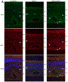
Immunohistochemistry of retinal sections shows IDO (red) partially colocalizing with CD-34 (green) in the capillary endothelium (arrowheads) of the retina of diabetic donors with DR (right). IDO-positive staining was also observed in the capillary endothelium of the retina of diabetic donors without retinopathy (middle) and nondiabetic donors (left), but the signal appears diffuse and less intense (arrowheads). Additionally, some of the IDO signal in the inner retina of diabetic donors with retinopathy appears not to colocalize with CD-34 (arrows), suggesting expression in glial cells. Nuclei are stained with DAPI (blue). (Adapted from Nahomi et al., 2018)
Figure 7.
Figure 7.. High levels of IFN-γ in the retina of diabetic donors with DR.
IFN-γ levels were measured by ELISA in retinal homogenates from age-matched nondiabetic (n = 9), diabetic (without retinopathy, n = 8), and diabetic (with retinopathy, n = 7) donors. Scale bar: 20 μm. *P < 0.05; NS, not significant. (Adapted from Nahomi et al., 2018)
Figure 8.
Figure 8.. Activation and self-perpetuation of the response of local immune competent cells in diabetic retinopathy.
Diabetes activates immune competent cells including micro- and macroglia (astrocytes and Müller glial cells) via multiple mechanisms, potentially inducing a self-perpeutating and amplification loop. Diabetes can directly induce activation of quiescent microglia (orange) leading to increase in number, change in morphology, and increased production of pro-inflammatory mediators. Similarly, diabetes also induces macroglia (green) to undergo activation and result in increased production of pro-inflammatory mediators. Finally, endothelial cells and neurons (pink) can also respond to diabetes by producing pro-inflammatory mediators. Any and all of these responses participate in the shift from immune-privileged to a highly pro-inflammatory ocular environment, and amplifies the activation of micro-and macroglia in an autocrine, and paracrine fashion. Once set in motion, this creates a loop in which de-activation of these immune competent cells is rendered highly difficult even when the trigger (i.e. suboptimal controlled diabetes) has been “removed”.
Figure 9.
Figure 9.. Complement system components are found in increasing levels in the neuroretina of donors with non-proliferative diabetic retinopathy compared to noon-diabetic donors (age, gender and race matched).
Immunohistochemical staining was performed on retinal sections of human donors without diabetes (Left) and with diabetes and non-proliferative diabetic retinopathy (NPDR, middle). The ganglion cell layer (GCL), inner nuclear layer (INL), and outer nuclear layer (ONL) are displayed in the first two panels. Agrin (red) a marker of blood vessels, and the complement system regulator CD59 (green) are shown with nuclei counterstain (blue). The right panel is a higher magnification image of the central panel demonstrating the presence CD59 away from agrin, in the neuroretina tissue.
Figure 10.
Figure 10.. Measurement of complement activation products C3a, C4a and C5a in human serum samples.
Levels of C3a (A) and C5a (B), but not C4a (C) were significantly higher in the DR group compared to age-matched healthy controls. Data were mean ± SD. Each dot represents one patient (Adapted from Lingjun Zhang et al., 2016).
Figure 11.
Figure 11.. C3 activation is important in paclitaxel-induced mechanical allodynia in CIPN.
A. Complement is activated after paclitaxel administration. WT or C3 KO rats were i.p. injected with DMSO (Vehicle) or 1 mg/kg paclitaxel (in DMSO according to the instructions of the manufacturer, Tocris, Bristol, UK) for 4 days (day 1 to 4), and then sera were collected on day 5 when the behavioral tests confirmed the development of mechanical allodynia and probed with an anti-C3 IgG to assess complement (C3) activation. There was a significant reduction of the α band (~130KDa) of C3 and an increased α2 chain of iC3b (~40 KDa) in the paclitaxel-treated WT rats. No C3 protein was detectable in sera from the C3 KO rats. B. C3 KO rats showed increased paw withdrawal threshold (PWT). WT and C3 KO rats (n=10/group) were injected with paclitaxel for 4 days (day 1 to 4), PWT was assessed on days 0, 7 and 11.; * p<0.05. (Adapted from Xu et al., 2018)
Figure 12.
Figure 12.. Summary of the clinical manifestations of diabetic retinopathy and the relative involvement of the vascular, immune, and neuronal systems.
Early on, activated micro- and macroglia (astrocytes and Muller glia) contribute to aneurysm formation through secretion of mediators that influence neighboring endothelial cells and weaken BRB integrity. Additionally, activated microglia secrete neurotoxins that impair the function of neurons in the neuroretina. As disease progress, activated microglia and macroglia contribute to the creation and formation of fibrovascular scars that push the disease stage to proliferative diabetic retinopathy. Activated microglia and astrocytes secrete pro-angiogenic and pro-inflammatory factors that result in the growth of abnormal vessels.

Similar articles

Cited by

References

    1. Abcouwer SF, Gardner TW, 2014. Diabetic retinopathy: Loss of neuroretinal adaptation to the diabetic metabolic environment. Ann. N. Y. Acad. Sci 1311, 174–190. 10.1111/nyas.12412 - DOI - PMC - PubMed
    1. Abhary S, Hewitt AW, Burdon KP, Craig JE, 2009. A systematic meta-analysis of genetic association studies for diabetic retinopathy. Diabetes 58, 2137–2147. 10.2337/db09-0059 - DOI - PMC - PubMed
    1. Abu El-Asrar AM, Dralands L, Missotten L, Al-Jadaan IA, Geboes K, 2004. Expression of apoptosis markers in the retinas of human subjects with diabetes. Investig. Ophthalmol. Vis. Sci 45, 2760–2766. 10.1167/iovs.03-1392 - DOI - PubMed
    1. Abukawa H, Tomi M, Kiyokawa J, Hori S, Kondo T, Terasaki T, Hosoya K-I, 2009. Modulation of retinal capillary endothelial cells by Müller glial cell-derived factors. Mol. Vis 15, 451–7. - PMC - PubMed
    1. Adams AJ, Bearse MA, 2012. Retinal neuropathy precedes vasculopathy in diabetes: A function-based opportunity for early treatment intervention? Clin. Exp. Optom 95, 256–265. 10.1111/j.1444-0938.2012.00733.x - DOI - PubMed

Publication types

Substances