

Review of Dermoscopy and Reflectance Confocal Microscopy Features of the Mucosal Melanoma
- PMID: 33429900
- PMCID: PMC7827612
- DOI: 10.3390/diagnostics11010091
Review of Dermoscopy and Reflectance Confocal Microscopy Features of the Mucosal Melanoma
Abstract
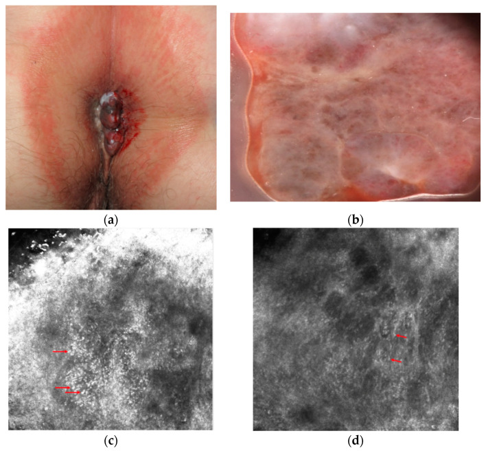
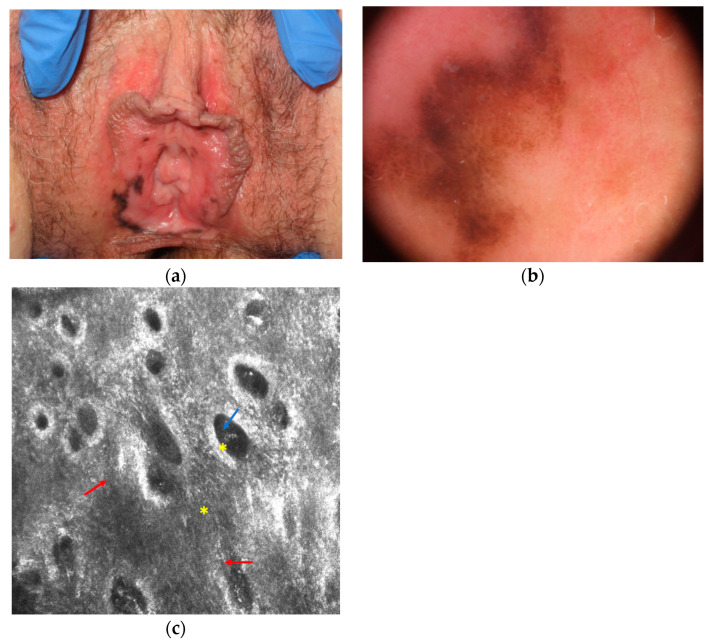
Mucosal melanoma is a rare tumor with aggressive biological behavior and poor prognosis. Diagnosis is often performed at an advanced stage when the lesions become symptomatic. Although dermoscopy and reflectance confocal microscopy (RCM) are widely used techniques for the diagnosis of cutaneous tumors, their use for mucosal lesions is not well established, probably because the latter are rarer. The objective of this study was to evaluate current literature on these imaging techniques for mucosal melanoma. We searched in PubMed and Cochrane databases all studies up to October 2020 dealing with dermoscopy, RCM, and mucosal melanoma. We found that the most relevant dermoscopic features were structureless pattern and/or the presence of multiple colors. RCM examination mainly showed numerous basal hyper-reflective dendritic cells and loss of normal architecture of the papillae of the lamina propria. Although diagnostic algorithms have been proposed for both techniques, the limit of these methods is the absence of large studies and of standardized and shared diagnostic criteria.
Keywords: dermoscopy; melanoma; mucosa; reflectance confocal microscopy.
Conflict of interest statement
The authors declare no conflict of interest.
Figures
Similar articles
-
Role of In Vivo Reflectance Confocal Microscopy in the Analysis of Melanocytic Lesions.Acta Dermatovenerol Croat. 2018 Apr;26(1):64-67. Acta Dermatovenerol Croat. 2018. PMID: 29782304 Review.
-
In Vivo Reflectance Confocal Microscopy for the Diagnosis of Melanoma and Melanotic Macules of the Lip.JAMA Dermatol. 2017 Sep 1;153(9):882-891. doi: 10.1001/jamadermatol.2017.0504. JAMA Dermatol. 2017. PMID: 28467525 Free PMC article.
-
Diagnostic accuracy of pigmented labial macules by in vivo reflectance confocal microscopy and correlation among techniques.J Am Acad Dermatol. 2021 Nov;85(5):1151-1160. doi: 10.1016/j.jaad.2020.02.067. Epub 2020 Mar 5. J Am Acad Dermatol. 2021. PMID: 32147389
-
Lesions Mimicking Melanoma at Dermoscopy Confirmed Basal Cell Carcinoma: Evaluation with Reflectance Confocal Microscopy.Dermatology. 2019;235(1):35-44. doi: 10.1159/000493727. Epub 2018 Nov 7. Dermatology. 2019. PMID: 30404078
-
Nipple and areola lesions: review of dermoscopy and reflectance confocal microscopy features.J Eur Acad Dermatol Venereol. 2019 Oct;33(10):1837-1846. doi: 10.1111/jdv.15727. Epub 2019 Jul 16. J Eur Acad Dermatol Venereol. 2019. PMID: 31166040 Review.
Cited by
-
Confocal Assessment of Pigmented-Mucosal Lesions: A Monocentric, Retrospective Evaluation of Lip and Genital Area.Dermatol Pract Concept. 2024 Jan 1;14(1):e2024028. doi: 10.5826/dpc.1401a28. Dermatol Pract Concept. 2024. PMID: 38364417 Free PMC article.
-
Dermoscopy in the diagnosis of cutaneous lymphoma (Review).Exp Ther Med. 2022 Jun;23(6):377. doi: 10.3892/etm.2022.11304. Epub 2022 Apr 7. Exp Ther Med. 2022. PMID: 35495594 Free PMC article. Review.
-
In Vivo Reflectance Confocal Microscopy Applied to Acral Melanocytic Lesions: A Systematic Review of the Literature.Diagnostics (Basel). 2024 Sep 25;14(19):2134. doi: 10.3390/diagnostics14192134. Diagnostics (Basel). 2024. PMID: 39410538 Free PMC article. Review.
-
Treatment outcomes of mucosal melanoma of head and neck: Efficacy of immune checkpoint inhibitors for advanced disease.Front Surg. 2022 Dec 26;9:1032626. doi: 10.3389/fsurg.2022.1032626. eCollection 2022. Front Surg. 2022. PMID: 37082097 Free PMC article.
References
-
- Cinotti E., Chevallier J., Labeille B., Cambazard F., Thomas L., Balme B., Leccia M.T., D’Incan M., Vercherin P., Douchet C., et al. Mucosal melanoma: Clinical, histological andc-kitgene mutational profile of 86 French cases. J. Eur. Acad. Dermatol. Venereol. 2017;31:1834–1840. doi: 10.1111/jdv.14353. - DOI - PubMed
-
- González-García R., Naval-Gías L., Martos P.L., Nam-Cha S.H., Rodríguez-Campo F.J., Muñoz-Guerra M.F., Sastre-Pérez J. Melanoma of the oral mucosa. Clinical cases and review of the literature. Med. Oral Patol. Oral Cir. Bucal. 2005;10:264–271. - PubMed
Publication types
LinkOut - more resources
Full Text Sources
Other Literature Sources
