Review
. 2021 Jan 8;10(1):104.
doi: 10.3390/cells10010104.

Cellular and Molecular Mechanisms of R/S-Roscovitine and CDKs Related Inhibition under Both Focal and Global Cerebral Ischemia: A Focus on Neurovascular Unit and Immune Cells

Affiliations
Review

Cellular and Molecular Mechanisms of R/S-Roscovitine and CDKs Related Inhibition under Both Focal and Global Cerebral Ischemia: A Focus on Neurovascular Unit and Immune Cells

Lucas Le Roy et al. Cells. .

Abstract

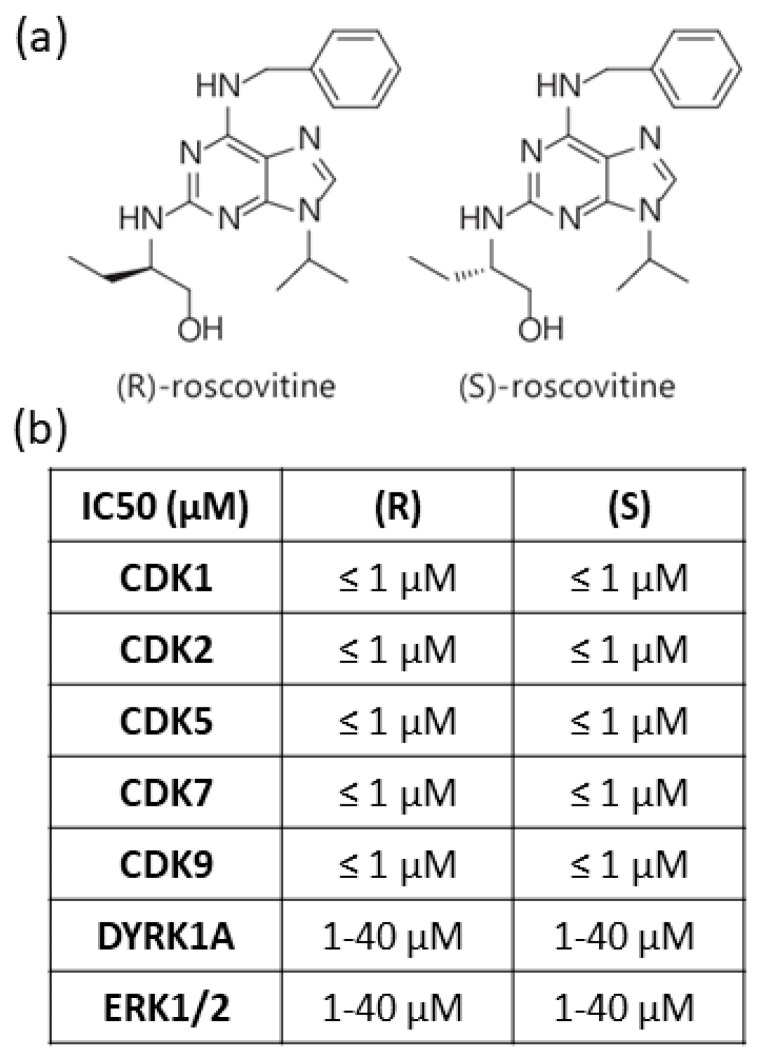
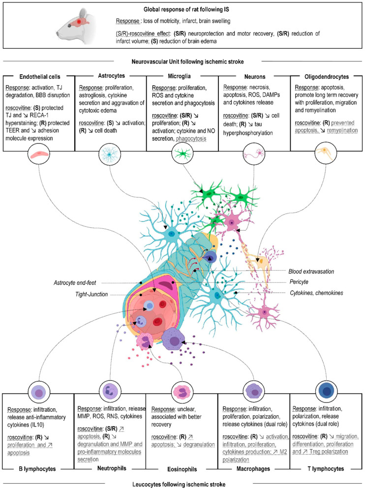
Ischemic stroke is the second leading cause of death worldwide. Following ischemic stroke, Neurovascular Unit (NVU) inflammation and peripheral leucocytes infiltration are major contributors to the extension of brain lesions. For a long time restricted to neurons, the 10 past years have shown the emergence of an increasing number of studies focusing on the role of Cyclin-Dependent Kinases (CDKs) on the other cells of NVU, as well as on the leucocytes. The most widely used CDKs inhibitor, (R)-roscovitine, and its (S) isomer both decreased brain lesions in models of global and focal cerebral ischemia. We previously showed that (S)-roscovitine acted, at least, by modulating NVU response to ischemia. Interestingly, roscovitine was shown to decrease leucocytes-mediated inflammation in several inflammatory models. Specific inhibition of roscovitine majors target CDK 1, 2, 5, 7, and 9 showed that these CDKs played key roles in inflammatory processes of NVU cells and leucocytes after brain lesions, including ischemic stroke. The data summarized here support the investigation of roscovitine as a potential therapeutic agent for the treatment of ischemic stroke, and provide an overview of CDK 1, 2, 5, 7, and 9 functions in brain cells and leucocytes during cerebral ischemia.

Keywords: CDK; ischemic stroke; leucocytes; neurovascular unit; roscovitine.


Conflict of interest statement

Serge Timsit is the co-inventor of a patent concerning (S)-roscovitine. The authors declare no conflict of interest.

Figures

Figure 1
Figure 1
(R)-roscovitine and (S)-roscovitine: structure and activity. (a) Structure of (R)- and (S)- stereoisomer of roscovitine. (b) IC50 values (in µM) described for these compounds.
Figure 2
Figure 2
(R) and (S)-roscovitine effect on Neurovascular Unit (NVU) and immune cells following ischemic stroke. A box was assigned to each cell from the NVU, in which two topics are described: the cell response after ischemic stroke, and the cellular effect of (R) and (S)-roscovitine. The box content is a summary of the data described for each cell type. For more detailed information, please refer to the full text of the manuscript. The (R)-roscovitine is represented by (R) and the (S)-roscovitine by (S). Black text means that the effect of roscovitine was observed in ischemic stroke model in vitro or in vivo. Grey text with dashed highlights means that the (R) or (S)-roscovitine effect was observed in other non-ischemic models, in vitro or in vivo. Created with BioRender.com.
Figure 3
Figure 3
Lymphocyte Th17/Treg balance. CD4+ T cells differentiate in Th1, Th2, Th17, or iTreg (induced regulatory T cells) depending on external stimuli. Th1 cells may aggravate brain injury by secreting pro-inflammatory cytokines (through T-bet), while Th2 cells may have neuroprotective effects on the injured brain by secreting anti-inflammatory cytokines (through GATA-3). The differentiation of Th17 cells from naïve T cells requires TGF-β and IL-6, which induce RORγt expression. Th17 cell is characterized by pro-inflammatory cytokines secretion, such as IL17A. In contrast, Foxp3, the master regulator of Tregs is induced by TGF-β and IL-2, and Treg secretes anti-inflammatory cytokines such as TGF-β and IL-10. Th17 and iTregs reciprocally inhibit their differentiation. Created with BioRender.com.

Similar articles

Cited by

References

    1. Moretti A., Ferrari F., Villa R.F. Neuroprotection for Ischaemic Stroke: Current Status and Challenges. Pharmacol. Ther. 2015;146:23–34. doi: 10.1016/j.pharmthera.2014.09.003. - DOI - PubMed
    1. Johnson C.O., Nguyen M., Roth G.A., Nichols E., Alam T., Abate D., Abd-Allah F., Abdelalim A., Abraha H.N., Abu-Rmeileh N.M., et al. Global, Regional, and National Burden of Stroke, 1990–2016: A Systematic Analysis for the Global Burden of Disease Study 2016. Lancet Neurol. 2019;18:439–458. doi: 10.1016/S1474-4422(19)30034-1. - DOI - PMC - PubMed
    1. Kaufmann A.M., Firlik A.D., Fukui M.B., Wechsler L.R., Jungries C.A., Yonas H. Ischemic Core and Penumbra in Human Stroke. Stroke. 1999;30:93–99. doi: 10.1161/01.STR.30.1.93. - DOI - PubMed
    1. Aggoun-Zouaoui D., Margalli I., Borrega F., Represa A., Plotkine M., Ben-Ari Y., Charriaut-Marlangue C. Ultrastructural Morphology of Neuronal Death Following Reversible Focal Ischemia in the Rat. Apoptosis. 1998;3:133–141. doi: 10.1023/A:1009653126347. - DOI - PubMed
    1. Astrup J., Symon L., Branston N.M., Lassen N.A. Cortical Evoked Potential and Extracellular K+ and H+ at Critical Levels of Brain Ischemia. Stroke. 1977;8:51–57. doi: 10.1161/01.STR.8.1.51. - DOI - PubMed

LinkOut - more resources