. 2021 Jan 7;13(2):182.
doi: 10.3390/cancers13020182.

Bromamine T (BAT) Exerts Stronger Anti-Cancer Properties than Taurine (Tau)

Affiliations

Bromamine T (BAT) Exerts Stronger Anti-Cancer Properties than Taurine (Tau)

Stella Baliou et al. Cancers (Basel). .

Abstract

Background: Taurine (Tau) ameliorates cancer pathogenesis. Researchers have focused on the functional properties of bromamine T (BAT), a stable active bromine molecule. Both N-bromotaurine (TauNHBr) and BAT exert potent anti-inflammatory properties, but the landscape remains obscure concerning the anti-cancer effect of BAT.

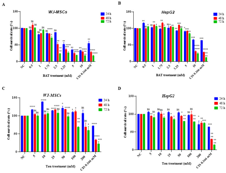
Methods: We used Crystal Violet, colony formation, flow cytometry and Western blot experiments to evaluate the effect of BAT and Tau on the apoptosis and autophagy of cancer cells. Xenograft experiments were used to determine the in vivo cytotoxicity of either agent.

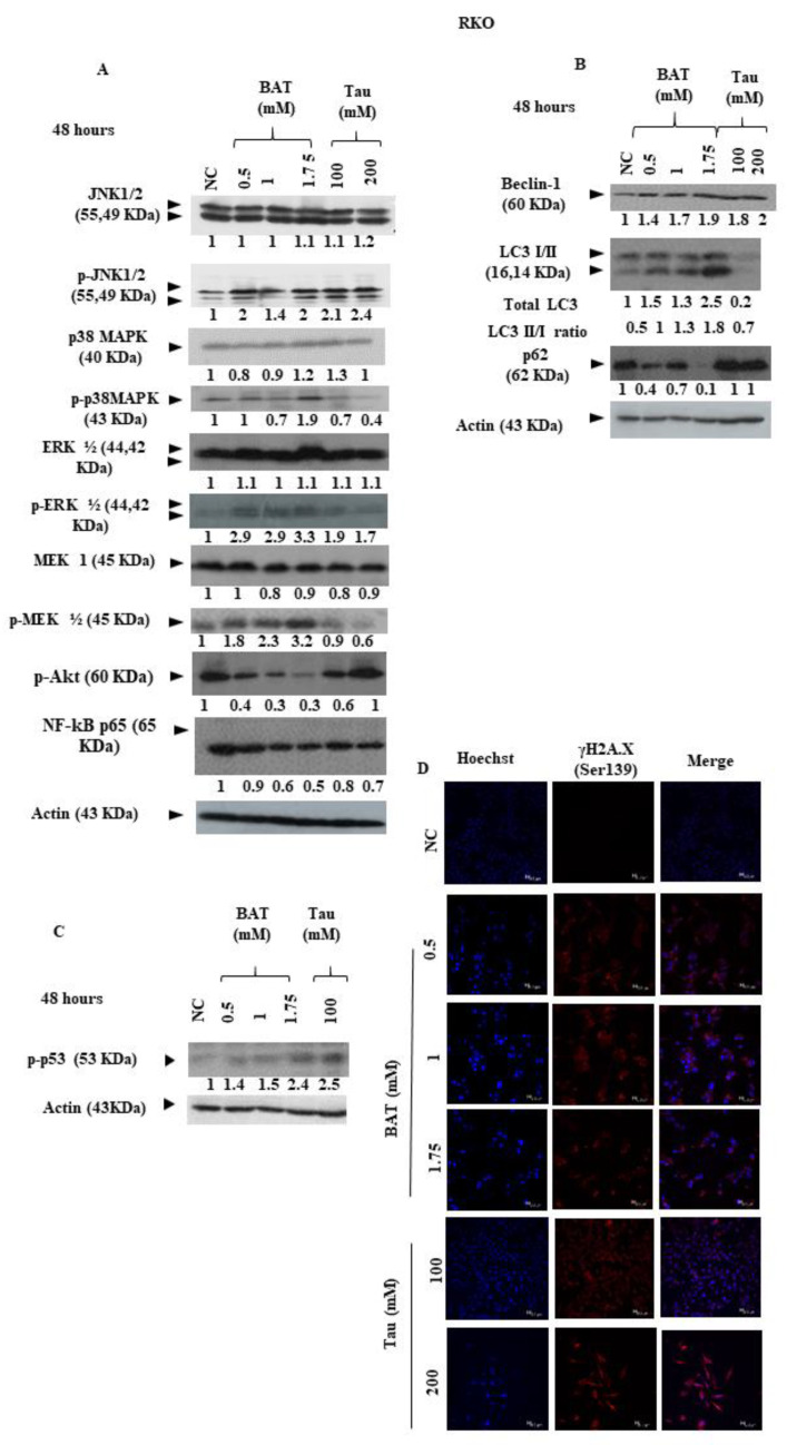
Results: We demonstrated that both BAT and Tau inhibited the growth of human colon, breast, cervical and skin cancer cell lines. Among them, BAT exerted the greatest cytotoxic effect on both RKO and MDA-MB-468 cells. In particular, BAT increased the phosphorylation of c-Jun N-terminal kinases (JNK½), p38 mitogen-activated protein kinase (MAPK), and extracellular-signal-regulated kinases (ERK½), thereby inducing mitochondrial apoptosis and autophagy in RKO cells. In contrast, Tau exerted its cytotoxic effect by upregulating JNK½ forms, thus triggering mitochondrial apoptosis in RKO cells. Accordingly, colon cancer growth was impaired in vivo.

Conclusions: BAT and Tau exerted their anti-tumor properties through the induction of (i) mitochondrial apoptosis, (ii) the MAPK family, and iii) autophagy, providing novel anti-cancer therapeutic modalities.

Keywords: breast cancer; bromamine T; colon cancer; taurine.


Conflict of interest statement

The authors declare no conflict of interest.

Figures

Figure 1
Figure 1
Bromamine T (BAT) is cytotoxic on a wide spectrum of cancer cells. The following cells: (A) RKO, (B) Caco2, (C) HT-29 (D) MDA-MB-231, (E) MDA-MB-468, (F) HeLa, (G) WM-164 cells were treated with (0.5–10 mΜ) BAT or 0.166 mM cisplatin (CIS) for 24–72 h. The percentage of viable cells was assessed upon BAT or Tau treatment versus negative control (NC) using the Crystal Violet procedure and statistical analysis was performed. ns: not significant, * p < 0.05. ** p < 0.01. *** p < 0.001.**** p < 0.0001.
Figure 2
Figure 2
Tau is cytotoxic on a wide spectrum of cancer cells. The following cells: (A) RKO, (B) Caco2, (C) HT-29 (D) MDA-MB-231, (E) MDA-MB-468, (F) HeLa, (G) WM-164 cells were treated with (5–200 mΜ) Tau or 0.166 mM CIS for 24–72 h. The percentage of viable cells upon BAT or Tau treatment versus negative control (NC) was assessed, using the Crystal Violet procedure and statistical analysis was performed. ns: not significant, * p < 0.05. ** p < 0.01. *** p < 0.001. **** p < 0.0001.
Figure 3
Figure 3
Both BAT and Tau exerted their cytotoxicity in a concentration-dependent manner. The following cells: (A,C) Wharton’s Jelly mesenchymal stem cells (WJ-MSCs) and (B,D) HepG2 cells were treated with (0.5–10 mΜ) BAT or (5–200 mΜ) Tau or 0.166 mM CIS for 24–72 h. The percentage of viable cells upon BAT or Tau treatment versus negative control (NC) was assessed, using the Crystal Violet procedure and statistical analysis was performed. ns: not significant, * p < 0.05. ** p < 0.01. *** p < 0.001. **** p < 0.0001.
Figure 4
Figure 4
Both BAT and Tau seem to have a growth-inhibitory effect on the colon, breast, and cervical cancer cell growth in an anchorage-independent manner. Clonogenic growth images of (A,B) RKO, (C,D) MDA-MB-468, and (E,F) HeLa cells treated with (0.5–1.75 mΜ) BAT or (100–200 mM) Tau were taken after 9 days (magnification ×100). The number of colonies that occupied the area of the plate was measured, using the Promega Cell counter software. Graphs (G,I,K) and (H,J,L) represent the quantitative and statistical analysis of colony formation assays, following BAT and Tau treatment versus the negative control (NC), respectively. ns: not significant, * p < 0.05. ** p < 0.01. *** p < 0.001. **** p < 0.0001.
Figure 5
Figure 5
BAT and Tau do not affect cancer cell cycle regulation: RKO (A,B,G,H), MDA-MB-468 (C,D,I,J), and HeLa cells (E,F,K,L) were treated with the indicated concentrations of BAT (AF) or Tau (GL) for 48 h in a cell-type-dependent manner. Cells were stained with propidium iodide (PI) and they were subjected to flow cytometry (FACS) analysis to determine the cell distributions in each phase of the cell cycle in the treated groups versus negative control (NC), using BD FACS Calibur and CellQuest Pro software. Graphs (A,C,E) and (G,I,K) show the percentage of apoptotic cells and statistical analysis compared to NC, following the treatment with BAT and Tau, respectively. Graphs (B,D,F) and (H,J,L) show the percentage of cells in each phase of cell cycle of non-apoptotic cells and statistical analysis compared to NC, upon the treatment with BAT and Tau, respectively. ns: not significant, * p < 0.05. ** p < 0.01. *** p < 0.001, **** p < 0.0001.
Figure 5
Figure 5
BAT and Tau do not affect cancer cell cycle regulation: RKO (A,B,G,H), MDA-MB-468 (C,D,I,J), and HeLa cells (E,F,K,L) were treated with the indicated concentrations of BAT (AF) or Tau (GL) for 48 h in a cell-type-dependent manner. Cells were stained with propidium iodide (PI) and they were subjected to flow cytometry (FACS) analysis to determine the cell distributions in each phase of the cell cycle in the treated groups versus negative control (NC), using BD FACS Calibur and CellQuest Pro software. Graphs (A,C,E) and (G,I,K) show the percentage of apoptotic cells and statistical analysis compared to NC, following the treatment with BAT and Tau, respectively. Graphs (B,D,F) and (H,J,L) show the percentage of cells in each phase of cell cycle of non-apoptotic cells and statistical analysis compared to NC, upon the treatment with BAT and Tau, respectively. ns: not significant, * p < 0.05. ** p < 0.01. *** p < 0.001, **** p < 0.0001.
Figure 6
Figure 6
Both BAT and Tau induce mitochondrial apoptotic cell death in cancer cells. (A) RKO, (B) MDA-MB-468 cells were treated with (0.5–1.75 mM) BAT or (100–200 mM) Tau for 48 h, versus the negative control (NC). The total protein expression levels of p53, Puma, Bak, Bax, Bim, Bik, p-Bad (Ser112), Bid, Bcl-xL, Bcl-2, total PARP, total caspase 3, cleaved caspase 3 and p21 were determined by immunoblotting. β-actin was used as a loading control. The relative intensity of each molecule was compared to that of NC. The whole gel figures are shown in Figure S3 while quantification and statistical analysis are shown in Figure S5 and Table S10. (C,D) Staining with CM-H2DCFDA dye demonstrated the oxidative burst in RKO or MDA-MB-468 cells after treatment with (0.5–1.75 mM) BAT or (100–200 mM) Tau for 48 h, versus the negative control (NC). ns: not significant, * p < 0.05.
Figure 6
Figure 6
Both BAT and Tau induce mitochondrial apoptotic cell death in cancer cells. (A) RKO, (B) MDA-MB-468 cells were treated with (0.5–1.75 mM) BAT or (100–200 mM) Tau for 48 h, versus the negative control (NC). The total protein expression levels of p53, Puma, Bak, Bax, Bim, Bik, p-Bad (Ser112), Bid, Bcl-xL, Bcl-2, total PARP, total caspase 3, cleaved caspase 3 and p21 were determined by immunoblotting. β-actin was used as a loading control. The relative intensity of each molecule was compared to that of NC. The whole gel figures are shown in Figure S3 while quantification and statistical analysis are shown in Figure S5 and Table S10. (C,D) Staining with CM-H2DCFDA dye demonstrated the oxidative burst in RKO or MDA-MB-468 cells after treatment with (0.5–1.75 mM) BAT or (100–200 mM) Tau for 48 h, versus the negative control (NC). ns: not significant, * p < 0.05.
Figure 7
Figure 7
Both BAT and Tau stimulate the mitochondrial apoptotic pathway in cancer cells: (A) immunofluorescence staining for cleaved caspase 3 (Asp175) in RKO cells after treatment with (0.5–1.75 mM) BAT or (100–200 mM) Tau for 48 h versus negative control (NC). Hoechst was used to stain the cell nuclei (magnification ×400); (B) immunofluorescence staining for cleaved caspase 3 (Asp175) in MDA-MB-468 cells after treatment with (0.5–1.75 mM) BAT or (100–200 mM) Tau for 48 h versus negative control (NC). Hoechst was used to stain the cell nuclei (magnification ×400). Quantification and statistical analysis are shown in Figure S6.
Figure 8
Figure 8
Both BAT and Tau affect the MAPK signaling pathway, autophagy, and DNA damage response (DDR) of RKO cells: (A) cells were treated with 0.5–1.75 mM BAT or 100–200 mM Tau for 48 h, versus negative control (NC). The total expression profiles of JNK½, p-JNK½ (Thr183/Tyr185), p38 MAPK, p-p38MAPK (Thr180/Tyr182), ERK½, p-ERK½ (Thr202/Tyr204), MEK-1, p-MEK½ (Ser217/Ser221), p-Akt (Ser473), NF-kB were determined by immunoblotting. β-actin was used as a loading control. The whole gel figures are shown in Figure S4 while quantification and statistical analysis are shown in Figure S7 and Table S10; (B) RKO cells were treated with (0.5–1.75 mM) BAT or (100–200 mM) Tau for 48 h, versus negative control (NC). The expression profiles of Beclin-1, LC3I/II, p62 were determined by immunoblotting. The whole gel figures are shown in Figure S4 while quantification and statistical analysis are shown in Figure S7 and Table S10. (C) RKO cells were treated with (0.5–1.75 mM) BAT or 100 mM Tau for 48 h, versus the negative control (NC). The expression profiles of p-p53 (Ser15) were determined by immunoblotting. β-actin was used as a loading control. The whole gel figures are shown in Figure S4 while quantification and statistical analysis are shown in Figure S7 and Table S10. (D) Immunofluorescence staining for γΗ2Α.X (Ser 139) after treatment with (0.5–1.75 mM) BAT or (100–200 mM) Tau for 48 h versus negative control (NC). Hoechst was used to stain the cell nuclei (magnification ×400). Quantification and statistical analysis are shown in Figure S7 and Table S10.
Figure 9
Figure 9
Both BAT and Tau mediate in vivo antitumor action in a xenograft model. (A) Schematic experimental design. A total of 1 × 10⁶ RKO cells were subcutaneously injected into the right/left flank of severe combined immune-deficient (SCID) mice (day −12). When the tumors became palpable, reaching the appropriate volume of 30–40 mm³ (day 1), the tumor-bearing mice were randomly assigned to 3 groups (6 mice/group). The first group was used as a negative control (NC) group, injected with phosphate buffer solution (PBS), and the other groups received an injection (3 mg/mouse, total 5 doses) of either agent directly into the tumor on specific days according to the timeline. Mice were sacrificed 28 days after the first day of tumor appearance (day 1). (B) Graph representation of the mean tumor volume in an approximately 28-day period. (C) Statistical analysis of the mean tumor volume between the BAT or Tau-treated groups and the negative control (NC) group. ns: not significant, * p < 0.05. **** p < 0.0001.
Figure 10
Figure 10
The scheme shows the underlying molecular mechanisms of BAT on colon cancer. BAT induces reactive oxygen species (ROS) accumulation, which in turn mediates mitochondrial apoptosis and autophagy. In BAT-treated RKO cells, mitochondrial apoptosis was induced through the upregulation of JNK½, p38 MAPK, ERK½ kinases, and autophagy was stimulated via the activation of JNK½ kinases as well as the downregulation of Akt.

Similar articles

Cited by

References

    1. Torre L.A., Bray F., Siegel R.L., Ferlay J., Lortet-Tieulent J., Jemal A. Global Cancer Statistics, 2012: Global Cancer Statistics, 2012. CA Cancer J. Clin. 2015;65:87–108. doi: 10.3322/caac.21262. - DOI - PubMed
    1. Davies H., Bignell G.R., Cox C., Stephens P., Edkins S., Clegg S., Teague J., Woffendin H., Garnett M.J., Bottomley W., et al. Mutations of the BRAF Gene in Human Cancer. Nature. 2002;417:949–954. doi: 10.1038/nature00766. - DOI - PubMed
    1. The Cancer Genome Atlas Network Comprehensive Molecular Characterization of Human Colon and Rectal Cancer. Nature. 2012;487:330–337. doi: 10.1038/nature11252. - DOI - PMC - PubMed
    1. Di Nicolantonio F., Martini M., Molinari F., Sartore-Bianchi A., Arena S., Saletti P., De Dosso S., Mazzucchelli L., Frattini M., Siena S., et al. Wild-Type BRAF Is Required for Response to Panitumumab or Cetuximab in Metastatic Colorectal Cancer. J. Clin. Oncol. 2008;26:5705–5712. doi: 10.1200/JCO.2008.18.0786. - DOI - PubMed
    1. Hyman D.M., Puzanov I., Subbiah V., Faris J.E., Chau I., Blay J.-Y., Wolf J., Raje N.S., Diamond E.L., Hollebecque A., et al. Vemurafenib in Multiple Nonmelanoma Cancers with BRAF V600 Mutations. N. Engl. J. Med. 2015;373:726–736. doi: 10.1056/NEJMoa1502309. - DOI - PMC - PubMed

LinkOut - more resources