. 2021 Jan 7;13(2):185.
doi: 10.3390/cancers13020185.

Prognostic Value and Function of KLF5 in Papillary Thyroid Cancer

Affiliations

Prognostic Value and Function of KLF5 in Papillary Thyroid Cancer

Poyil Pratheeshkumar et al. Cancers (Basel). .

Abstract

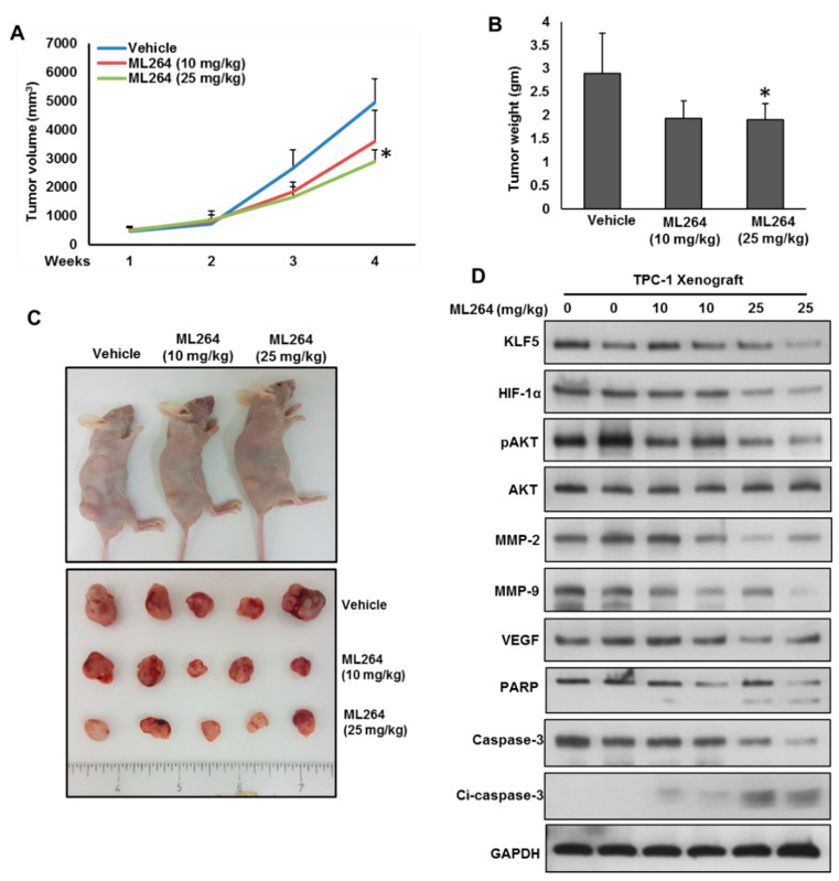
The Krüppel-like factor 5 (KLF5), a zinc-finger transcriptional factor, is highly expressed in several solid tumors, but its role in PTC remains unclear. We investigated the expression of KLF5 protein in a large cohort of PTC patient samples and explored its functional role and mechanism in PTC cell lines in vitro and in vivo. KLF5 overexpression was observed in 65.1% of all PTC cases and it was significantly associated with aggressive clinico-pathological parameters and poor outcome. Given the significant association between KLF5 and HIF-1α overexpression in PTC patients, we investigated the functional correlation between KLF5 and HIF-1α in PTC cells. Indeed, the analysis revealed the co-immunoprecipitation of KLF5 with HIF-1α in PTC cells. We also identified KLF5-binding sites in the HIF-1α promoter that specifically bound to KLF5 protein. Mechanistically, KLF5 promoted PTC cell growth, invasion, migration, and angiogenesis, while KLF5 downregulation via specific inhibitor or siRNA reverses its action in vitro. Importantly, the silencing of KLF5 decreases the self-renewal ability of spheroids generated from PTC cells. In addition, the depletion of KLF5 reduces PTC xenograft growth in vivo. These findings suggest KLF5 can be a possible new molecular therapeutic target for a subset of PTC.

Keywords: HIF-1α; KLF5; apoptosis; invasion; papillary thyroid cancer; stemness.


Conflict of interest statement

The authors declare no conflict of interest.

Figures

Figure 1
Figure 1
Immunohistochemical analysis of KLF5 and HIF-1α expression in Papillary Thyroid Cancer (PTC) TMA. (A) KLF5 expression in normal thyroid and papillary thyroid carcinoma (PTC) tissues. A significant difference in expression levels was noted between normal thyroid (n = 225) and PTC tissues (n = 1219) (p < 0.001). (B) PTC array spot showing overexpression of KLF5 (a) and HIF-1α (c). In contrast, another PTC tissue array spot showing low expression of KLF5 (b) and HIF-1α (d). 20X/0.70 objective on an Olympus BX 51 microscope. (Olympus America Inc, Center Valley, PA, USA) with the inset showing a 40X 0.85 aperture magnified view of the same TMA spot. (C) Kaplan–Meier survival analysis for the prognostic significance of KLF5 expression in PTC. PTC patients with overexpression of KLF5 had reduced disease-free survival at 5 years on univariate analysis compared to tumors showing low expression of KLF5 (p = 0.0066).
Figure 2
Figure 2
KLF5 is a functional upstream of HIF-1α. (A) Basal expression of KLF5 and HIF-1α in PTC cell lines. Proteins were isolated from three PTC cell lines and immunoblotted with antibodies against KLF5, HIF-1α and GAPDH. (B) KLF5 interact with HIF-1α. Cell lysates extracted from PTC cells were immunoprecipitated with KLF5 or IgG antibody. Interaction of endogenous KLF5 and HIF-1α was detected by immunoblotting. (C) HIF-1α interact with KLF5. Cell lysates extracted from PTC cells were immunoprecipitated with HIF-1α or IgG antibody. Interaction of endogenous KLF5 and HIF-1α was detected by immunoblotting. (D) ML264 disrupts the physical interaction of KLF5 and HIF-1α in PTC cells. PTC cells were treated with indicated doses of ML264 for 6 h. Cell lysates extracted from PTC cells were immunoprecipitated with KLF5 antibody. Interaction of endogenous KLF5 and HIF-1α was detected by immunoblotting. (E,F) KLF5 binding to HIF-1α promoter. For the ChIP assay, the KLF5-binding regions on HIF-1α promoter were identified. PTC cells were treated with and without ML264 (5 and 10 μm) for 6 h, fixed with formaldehyde, and cross-linked, and then chromatin was isolated. The chromatin was immunoprecipitated (IP) with an anti-KLF5 antibody or control mouse IgG. The KLF5 binding to the HIF-1α promoters was analyzed by regular PCR (E) or quantitative real-time PCR (F) with a primer specific to the KLF5-binding regions in HIF-1α promoter. The data represent the percent input and are normalized to each control. GAPDH was used as a loading control. (G) Silencing of KLF5 inhibits HIF-1α. PTC cells were transfected with scrambled siRNA and KLF5 siRNA (50 and 100 nM). After 48 h, cells were lysed and proteins were immunoblotted with antibodies against KLF5, HIF-1α and GAPDH. (H) ML264 treatment down-regulates the expression of KLF5 and HIF-1α in PTC cells. PTC cells were treated with indicated doses of ML264 for 48 h. After cell lysis, equal amounts of proteins were separated by SDS-PAGE, transferred to immobilon membrane, and immuno-blotted with antibodies against KLF5, HIF-1α and GAPDH as indicated. (I) Knockdown of HIF-1α has no effect on KLF5 expression. PTC cells were transfected with scrambled siRNA and HIF-1α siRNA (50 and 100 nM). After 48 h, cells were lysedand proteins were immunoblotted with antibodies against HIF-1α, KLF5 and GAPDH. (J) Forced expression of KLF5 increases HIF-1α expression. K1 cells were transfected with either empty vector or KLF5 cDNA for 48 h. Proteins were isolated and immunoblotted with antibodies against KLF5, HIF-1α and GAPDH for equal loading. Data presented in the bar graphs are the mean ± SD of triplicates in an independent experiments, which was repeated for at least two times with the same results. * Indicates a statistically significant difference compared to control with p < 0.05. Western blot experiments were repeated at least two times with the same results.
Figure 3
Figure 3
Downregulation of KLF5 inhibits tumor cell invasion, migration and angiogenesis. (A,B) KLF5 inhibition decreases the invasive capacity of PTC cells. PTC cells were treated with indicated doses ML264 and seeded into the upper compartment of invasion chambers. The bottom chambers were filled with RPMI media. After 24 h incubation, invaded cells were fixed, stained and quantified. (C) Silencing of KLF5 decreases invasion of PTC cells. PTC cells were transfected with scrambled siRNA and KLF5 siRNA (50 and 100 nM). After 48 h, cells were seeded into the upper compartment of invasion chambers. The bottom chambers were filled with RPMI media. After 24 h incubation, invaded cells were fixed, stained and quantified. (D) KLF5 inhibition causes reduction in the migration capacity of PTC cells. PTC cells were treated with indicated doses ML264 and seeded into the upper compartment of migration chambers. The bottom chambers were filled with RPMI media. After 24 h incubation, migrated cells were fixed, stained and quantified. (E) Silencing of KLF5 decreases migration of PTC cells. PTC cells were transfected with scrambled siRNA and KLF5 siRNA (50 and 100 nM). After 48 h, cells were seeded into the upper compartment of migration chambers. The bottom chambers were filled with RPMI media. After 24 h incubation, migrated cells were fixed, stained and quantified. (F) ML264 treatment down-regulates the expression of MMP-2, MMP-9 and VEGF in PTC cells. PTC cells were treated with indicated doses of ML264 for 48 h. After cell lysis, equal amounts of proteins were separated by SDS-PAGE, transferred to immobilon membrane, and immuno-blotted with antibodies against KLF5, HIF-1α, MMP-2, MMP-9, VEGF and GAPDH, as indicated. (G) Silencing of KLF5 down-regulates the expression of MMP-2, MMP-9 and VEGF in PTC cells. PTC cells were transfected with scrambled siRNA and KLF5 siRNA (50 and 100 nM). After 48 h, cells were lysed and proteins were immunoblotted with antibodies against KLF5, HIF-1α, MMP-2, MMP-9, VEGF and GAPDH. (H,I) KLF5 inhibition decreases HUVECs tube formation. HUVECs grown on matrigel were treated with conditioned media from KLF5 treated and untreated PTCs for 24 h, cells were fixed, and tubular structures were photographed and quantified. Data presented in the bar graphs are the mean ± SD of triplicates in an independent experiment which was repeated for at least two times with the same results. * Indicates a statistically significant difference compared to control with p < 0.05. Western blot experiments were repeated at least two times with the same results.
Figure 4
Figure 4
Downregulation of KLF5 inhibits PTC cell growth in vitro. (A) ML264 inhibits cell viability. PTC cells (104) were incubated with indicated doses of ML264 for 48 h. Cell viability was performed using MTT. (B,C) ML264 inhibited clonogenicity. PTC cells (8 × 10²) after ML264 treatment were seeded into two dishes (60 mm diameter), and grown for an additional 10 days, then stained with crystal violet, and colonies were counted. (D) Knockdown of KLF5 decreases clonogenicity. PTC cells were transfected with scrambled siRNA and KLF5 siRNA (50 and 100 nM). After 48 h, cells (8 × 10²) were seeded into two dishes (60 mm diameter), and grown for an additional 10 days, then stained with crystal violet, and colonies were counted. (E) ML264 induces apoptosis in PTC cell lines. PTC cells were treated with indicated doses of ML264 for 48 h and cells were stained with fluorescein-conjugated annexin-V and propidium iodide (PI) and analyzed by flow cytometry. (F) ML264 treatment causes inactivation of AKT and down-regulates the expression of anti-apoptotic proteins and induces the cleavage of caspase-3 and PARP. PTC cells were treated with indicated doses of ML264 for 48 h. After cell lysis, equal amounts of proteins were separated by SDS-PAGE, transferred to immobilon membrane, and immuno-blotted with antibodies against KLF5, HIF-1α, pAKT, AKT, Bcl-2, Bcl-xL, PARP, casapse-3, cleaved casapse-3 and GAPDH as indicated. All the experiments were repeated twice with similar results. Data presented in the bar graphs are the mean ± SD of triplicates in an independent experiment which was repeated at least two times with the same results. * Indicates a statistically significant difference compared to control with p < 0.05. Western blot experiments were repeated at least two times with the same results.
Figure 5
Figure 5
Inhibition of KLF5 decreases the self-renewal ability of spheroids generated from PTC cells. (A,B) Isolation of spheroid-forming cells from PTC cells. Sphere forming assay was performed by culturing PTC cells (5 × 10² cells/well) in sphere medium for 14 days in 24-well ultra-low attachment plates. Proteins were isolated from spheroid-forming cells and respective parental adherent cells and immunoblotted with antibodies against KLF5, HIF-1α, CD44, CD133, NANOG, OCT4 and GAPDH (A). Spheroid-forming cells and adherent cells were labelled with Aldefluor with and without ALDH inhibitor, DEAB and analyzed by flow cytometer according to the manufacturer’s instructions (B). (C,D) Silencing of KLF5 inhibits self-renewal ability of spheroids. PTC cells were transfected with KLF5 shRNA and cells were subjected to sphere forming assay. Spheroids in the entire well were counted. (E,F) Silencing of KLF5 inhibits stemness of spheroids as confirmed by immunoblotting using stem cell markers. PTC cells were transfected with scramble or KLF5 shRNA’s and grown in sphere medium. Proteins were isolated from spheroids and immunoblotted with antibodies against KLF5, HIF-1α, CD44, CD133, NANOG, OCT4 and GAPDH (E). ALDH activity was also determined (F). Data presented in the bar graphs are the mean ± SD of triplicates in an independent experiment which was repeated at least two times with the same results. * Indicates a statistically significant difference compared to control with p < 0.05. Western blot experiments were repeated at least two times with the same results.
Figure 6
Figure 6
Downregulation of KLF5 inhibited PTC cell growth in vivo. TPC-1 cells were subcutaneously injected into the flanks of 6-week old nude mice (4 × 10⁶ cells per mouse). After tumors grew to about 100 mm3, mice were treated intraperitoneally with indicated dose of ML264, twice a week for 30 days. (A) The volume of each tumor was measured every week. The average (n = 6) tumor volume in each group of mice was calculated. (B) After four weeks treatment, mice were sacrificed and mean tumor weight (±SD) was calculated in each group, * p < 0.05. (C) Representative tumor images of each group of mice. (D) Tissue lysates from tumors were immuno-blotted with antibodies against KLF5, HIF-1α, pAKT, AKT, MMP-2, MMP-9, VEGF, PARP, caspase-3, cleaved caspase-3 and GAPDH.

Similar articles

Cited by

References

    1. Howlader N., Noone A., Krapcho M., Neyman N., Aminou R., Altekruse S., Kosary C., Ruhl J., Tatalovich Z., Cho H. SEER Cancer Statistics Review, 1975–2009 (Vintage 2009 Populations) National Cancer Institute; Bethesda, MD, USA: 2012.
    1. Tumino D., Frasca F., Newbold K. Updates on the management of advanced, metastatic, and radioiodine refractory differentiated thyroid cancer. Front. Endocrinol. (Lausanne) 2017;8:312. doi: 10.3389/fendo.2017.00312. - DOI - PMC - PubMed
    1. Grant C.S. Recurrence of papillary thyroid cancer after optimized surgery. Gland Surg. 2015;4:52. - PMC - PubMed
    1. Lee J.-W., Bae S.-H., Jeong J.-W., Kim S.-H., Kim K.-W. Hypoxia-inducible factor (HIF-1) α: Its protein stability and biological functions. Exp. Mol. Med. 2004;36:1–12. doi: 10.1038/emm.2004.1. - DOI - PubMed
    1. Mori A., Moser C., Lang S.A., Hackl C., Gottfried E., Kreutz M., Schlitt H.J., Geissler E.K., Stoeltzing O. Up-regulation of Krüppel-like factor 5 in pancreatic cancer is promoted by interleukin-1β signaling and hypoxia-inducible factor-1α. Mol. Cancer Res. 2009;7:1390–1398. doi: 10.1158/1541-7786.MCR-08-0525. - DOI - PubMed

LinkOut - more resources