Common Mechanism for Target Specificity of Protein- and DNA-Targeting ADP-Ribosyltransferases
- PMID: 33430384
- PMCID: PMC7827354
- DOI: 10.3390/toxins13010040
Common Mechanism for Target Specificity of Protein- and DNA-Targeting ADP-Ribosyltransferases
Abstract
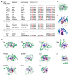
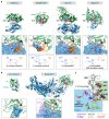
Many bacterial pathogens utilize ADP-ribosyltransferases (ARTs) as virulence factors. The critical aspect of ARTs is their target specificity. Each individual ART modifies a specific residue of its substrates, which could be proteins, DNA, or antibiotics. However, the mechanism underlying this specificity is poorly understood. Here, we review the substrate recognition mechanism and target residue specificity based on the available complex structures of ARTs and their substrates. We show that there are common mechanisms of target residue specificity among protein- and DNA-targeting ARTs.
Keywords: ADP-ribosyltransferase; complex structure of enzyme and substrate; substrate recognition; target residue specificity.
Conflict of interest statement
The authors declare no conflict of interest.
Figures


Similar articles
-
Reaction Mechanism of Mono-ADP-Ribosyltransferase Based on Structures of the Complex of Enzyme and Substrate Protein.Curr Top Microbiol Immunol. 2015;384:69-87. doi: 10.1007/82_2014_415. Curr Top Microbiol Immunol. 2015. PMID: 24990621 Review.
-
General ADP-Ribosylation Mechanism Based on the Structure of ADP-Ribosyltransferase-Substrate Complexes.Toxins (Basel). 2024 Jul 11;16(7):313. doi: 10.3390/toxins16070313. Toxins (Basel). 2024. PMID: 39057953 Free PMC article. Review.
-
The family of toxin-related ecto-ADP-ribosyltransferases in humans and the mouse.Protein Sci. 2002 Jul;11(7):1657-70. doi: 10.1110/ps.0200602. Protein Sci. 2002. PMID: 12070318 Free PMC article.
-
Substrate N2 atom recognition mechanism in pierisin family DNA-targeting, guanine-specific ADP-ribosyltransferase ScARP.J Biol Chem. 2018 Sep 7;293(36):13768-13774. doi: 10.1074/jbc.AC118.004412. Epub 2018 Aug 2. J Biol Chem. 2018. PMID: 30072382 Free PMC article.
-
Ecto-ADP-ribosyltransferases (ARTs): emerging actors in cell communication and signaling.Curr Med Chem. 2004 Apr;11(7):857-72. doi: 10.2174/0929867043455611. Curr Med Chem. 2004. PMID: 15078170 Review.
Cited by
-
Mechanism of threonine ADP-ribosylation of F-actin by a Tc toxin.Nat Commun. 2022 Jul 20;13(1):4202. doi: 10.1038/s41467-022-31836-w. Nat Commun. 2022. PMID: 35858890 Free PMC article.
-
Genomic epidemiology of rifampicin ADP-ribosyltransferase (Arr) in the Bacteria domain.Sci Rep. 2021 Oct 5;11(1):19775. doi: 10.1038/s41598-021-99255-3. Sci Rep. 2021. PMID: 34611248 Free PMC article.
-
ADP-ribosylation systems in bacteria and viruses.Comput Struct Biotechnol J. 2021 Apr 17;19:2366-2383. doi: 10.1016/j.csbj.2021.04.023. eCollection 2021. Comput Struct Biotechnol J. 2021. PMID: 34025930 Free PMC article. Review.
-
Molecular determinants of nucleic acid recognition by an RNA-targeting ADP-ribosyltransferase toxin.J Biol Chem. 2025 Jul 7;301(8):110463. doi: 10.1016/j.jbc.2025.110463. Online ahead of print. J Biol Chem. 2025. PMID: 40633614 Free PMC article.
-
Venomous Cargo: Diverse Toxin-Related Proteins Are Associated with Extracellular Vesicles in Parasitoid Wasp Venom.Pathogens. 2025 Mar 5;14(3):255. doi: 10.3390/pathogens14030255. Pathogens. 2025. PMID: 40137740 Free PMC article.
References
Publication types
MeSH terms
Substances
Grants and funding
- 15K08289/Japan Society for the Promotion of Science/International
- 17K15095/Japan Society for the Promotion of Science/International
- 23121529/Ministry of Education, Culture, Sports, Science and Technology/International
- 25121733/Ministry of Education, Culture, Sports, Science and Technology/International
LinkOut - more resources
Full Text Sources
Other Literature Sources

