Review
. 2021 Jan 7;10(1):94.
doi: 10.3390/cells10010094.

Context Matters: NOTCH Signatures and Pathway in Cancer Progression and Metastasis

Affiliations
Review

Context Matters: NOTCH Signatures and Pathway in Cancer Progression and Metastasis

Julia O Misiorek et al. Cells. .

Abstract

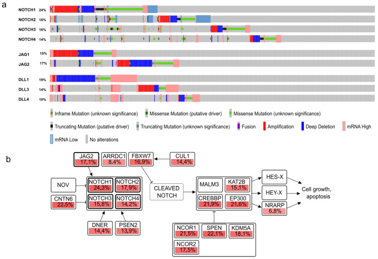
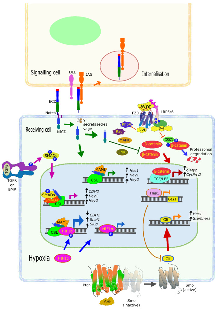
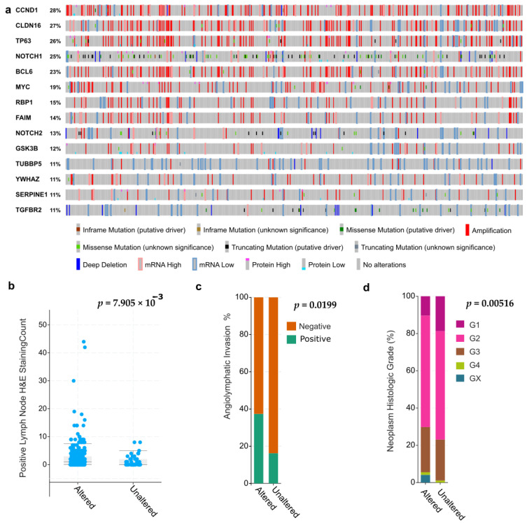
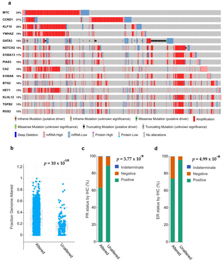
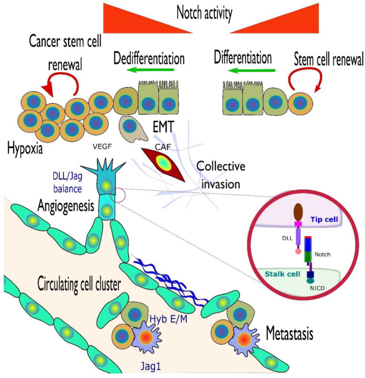
The Notch signaling pathway is a critical player in embryogenesis but also plays various roles in tumorigenesis, with both tumor suppressor and oncogenic activities. Mutations, deletions, amplifications, or over-expression of Notch receptors, ligands, and a growing list of downstream Notch-activated genes have by now been described for most human cancer types. Yet, it often remains unclear what may be the functional impact of these changes for tumor biology, initiation, and progression, for cancer therapy, and for personalized medicine. Emerging data indicate that Notch signaling can also contribute to increased aggressive properties such as invasion, tumor heterogeneity, angiogenesis, or tumor cell dormancy within solid cancer tissues; especially in epithelial cancers, which are in the center of this review. Notch further supports the "stemness" of cancer cells and helps define the stem cell niche for their long-term survival, by integrating the interaction between cancer cells and the cells of the tumor microenvironment (TME). The complexity of Notch crosstalk with other signaling pathways and its roles in cell fate and trans-differentiation processes such as epithelial-to-mesenchymal transition (EMT) point to this pathway as a decisive player that may tip the balance between tumor suppression and promotion, differentiation and invasion. Here we not only review the literature, but also explore genomic databases with a specific focus on Notch signatures, and how they relate to different stages in tumor development. Altered Notch signaling hereby plays a key role for tumor cell survival and coping with a broad spectrum of vital issues, contributing to failed therapies, poor patient outcome, and loss of lives.

Keywords: Notch signaling pathway; angiogenesis; epithelial-mesenchymal transition (EMT); gain and loss of function mutations (GOF and LOF); metastasis; oncogenic mutations; personalized cancer medicine; tumor microenvironment (TME); tumor progression; tumor suppressor gene.


Conflict of interest statement

The authors declare no conflict of interest.

Figures

Figure 1
Figure 1
Genetic alterations in Notch pathway genes across human cancer cell lines. (a) Mutual exclusivity of genetic events targeting NOTCH receptors and ligands across 1000+ cell lines from the Cancer Cell Line Encyclopedia (CCLE) collection, as displayed in the cBioPortal genome browser. The sorting by mutation type illustrates that genetic mutations affect typically only one of the four NOTCH receptors. Similar mutual exclusivity is observed for the JAG1 and 2, and DLL1, DLL3, and DLL4 ligands. (b) Percentage of genetic events (mutations, amplifications, deletions, over-expression) observed in core genes of the Notch pathway, across the 1000+ cell lines of the CCLE collection/BROAD Institute (https://www.cbioportal.org/study?id=ccle_broad_2019). Gene Symbols as identifiers used according to Genome Reference Consortium Human Build 37 (GRCh37).
Figure 2
Figure 2
Notch crosstalk with other signaling pathways. Activation of Notch receptors by ligand-presenting cells results in the release of the Notch intracellular domain (NICD), forming a transcriptional activator complex with CBF1, Suppressor of Hairless, Lag-1 (CSL) and mastermind-like protein 1 (MAML). It also interacts with other signaling pathways, for example, by binding to β-catenin and inducing its degradation. The Wnt/β-catenin pathway is involved in cell cycle regulation. NICD also interacts with hypoxia-inducible factor 1-alpha (HIF1a) during hypoxia enhancing HIF1a transactivating activity. Likewise, SMAD transcription factors, which are activated downstream the TGFR and BMP pathways, also interact with NICD/CSL complex. Interactions with additional pathways include communication with Sonic Hedgehog (SHH), where binding to Patched (Ptch) results in the release of Smoothened (Smo) and activation of a cascade of signaling centered in Gli. Hes1, a downstream target of Notch, represses GLI, while Gli activation by Smo triggers downstream genes such as HES1 and other stemness genes. Gene symbols used according to Genome Reference Consortium Human Build 37 (GRCh37).
Figure 3
Figure 3
Analysis of most frequently altered NOTCH and EMT-related genes in head and neck squamous cell carcinomas (HNSCCC). (a) Mutation and gene expression profiles for 12 target genes associated with both Notch signaling and EMT in the cBioPortal genomic database (sorted by patient number, not mutations). Indicated are mainly genomic amplifications, deletions, and point mutations, followed by mRNA and protein overexpression across 530 HNSCC tumor samples from the “Firehose legacy” sequencing project. (b) Number of patients with positive lymph nodes (left) is increased for tumors that harbor genetic alterations in the 12 Notch/EMT genes show above. (c) Patients with a high degree of genetic changes in Notch/EMT signature genes also show a larger proportion of tumors with angiolymphatic invasion, or penetration of tumor cells into lymph vessels in the tumor periphery. (d) Patients with large numbers of Notch/EMT genes mutated further show a larger likelihood to develop high grade tumors (G3 and G4), with enhanced invasive properties.
Figure 4
Figure 4
Analysis of the 12 most frequently mutated genes associated simultaneously with Notch signaling and EMT, in 1039 breast cancers (TCGA sequencing project). In contrast to Figure 3, tumors have been sorted according to most frequent mutations (CCND1, MYC) to illustrate the high frequency of amplifications. (a) Sorted incidence of mutations, including amplifications, deletions, point mutations, and RNA overexpression, as generated by the cBioPortal browser. Data have been sorted according to type of mutation/genetic alteration. (b) Tumors with high frequency of genetic alterations in Notch/EMT target or signature genes also show a higher degree of genetic instability. (c) Tumors with high levels of Notch/EMT-related genetic alterations more frequently belong to the triple-negative and basal-like breast cancers that lack expression of progesterone receptor (PR). (d) Corresponding finding for expression of estrogen receptor (ER) in the same tumors, as analyzed by immunohistochemistry (IHC).
Figure 5
Figure 5
Notch activity in different contexts, exemplified here within the context of epithelial tissues and cells (olive-green cells, partly with brush border). Notch activity is important in stem cell renewal and differentiation (top-right). This is hijacked in some cancer cell types to maintain stem-like features such as unlimited proliferation and acquiring resistance to radio/chemotherapy (cells orange-brown color), in others to gain a mesenchymal phenotype (gray cells) that can resemble the phenotype of cancer-associated fibroblasts (CAFs, dark brown) and may result in collective invasion, characteristic for epithelial cells. As tumors grow, they may become increasingly hypoxic, which triggers neo-angiogenesis. The vasculature (endothelial cells, turquoise) is positive for Notch receptors and ligands, thus the interaction of the vasculature with tumor cells is likely a factor in invasiveness. One of the most striking features of Notch signaling, via Jag1 and hyb-E/M (hybrid epithelial-to-mesenchymal transition), is its supportive role in circulating clusters of tumor cells that have particularly high metastatic potential (olive colored, rounded cells), partnering also with immune cells (stellate, blue cells with red nuclei). The inset shows the ligand–receptor activation between 2 cells. Gene symbols explained in the text.

Similar articles

Cited by

References

    1. Kopan R., Ilagan M.X.G. The canonical Notch signaling pathway: Unfolding the activation mechanism. Cell. 2009;137:216–233. doi: 10.1016/j.cell.2009.03.045. - DOI - PMC - PubMed
    1. Wu Z.Q., Rowe R.G., Lim K.C., Lin Y., Willis A., Tang Y., Li X.Y., Nor J.E., Maillard I., Weiss S.J. A Snail1/Notch1 signalling axis controls embryonic vascular development. Nat. Commun. 2014;5:1–15. doi: 10.1038/ncomms4998. - DOI - PMC - PubMed
    1. Hellström M., Phng L.K., Hofmann J.J., Wallgard E., Coultas L., Lindblom P., Alva J., Nilsson A.K., Karlsson L., Gaiano N., et al. Dll4 signalling through Notch1 regulates formation of tip cells during angiogenesis. Nature. 2007;445:776–780. doi: 10.1038/nature05571. - DOI - PubMed
    1. High F.A., Min M.L., Pear W.S., Loomes K.M., Kaestner K.H., Epstein J.A. Endothelial expression of the Notch ligand Jagged1 is required for vascular smooth muscle development. Proc. Natl. Acad. Sci. USA. 2008;105:1955–1959. doi: 10.1073/pnas.0709663105. - DOI - PMC - PubMed
    1. Boareto M., Jolly M.K., Ben-Jacob E., Onuchic J.N. Jagged mediates differences in normal and tumor angiogenesis by affecting tip-stalk fate decision. Proc. Natl. Acad. Sci. USA. 2015;112:E3836–E3844. doi: 10.1073/pnas.1511814112. - DOI - PMC - PubMed

Publication types

Substances

LinkOut - more resources