. 2021 Jan 7;10(1):93.
doi: 10.3390/cells10010093.

Acute D-Serine Co-Agonism of β-Cell NMDA Receptors Potentiates Glucose-Stimulated Insulin Secretion and Excitatory β-Cell Membrane Activity

Affiliations

Acute D-Serine Co-Agonism of β-Cell NMDA Receptors Potentiates Glucose-Stimulated Insulin Secretion and Excitatory β-Cell Membrane Activity

Amber Lockridge et al. Cells. .

Abstract

Insulin-secreting pancreatic β-cells express proteins characteristic of D-serine regulated synapses, but the acute effect of D-serine co-agonism on its presumptive β-cell target, N-methyl D-aspartate receptors (NMDARs), is unclear. We used multiple models to evaluate glucose homeostasis and insulin secretion in mice with a systemic increase in D-serine (intraperitoneal injection or DAAO mutants without D-serine catabolism) or tissue-specific loss of Grin1-encoded GluN1, the D-serine binding NMDAR subunit. We also investigated the effects of D-serine ± NMDA on glucose-stimulated insulin secretion (GSIS) and β-cell depolarizing membrane oscillations, using perforated patch electrophysiology, in β-cell-containing primary isolated mouse islets. In vivo models of elevated D-serine correlated to improved blood glucose and insulin levels. In vitro, D-serine potentiated GSIS and β-cell membrane excitation, dependent on NMDAR activating conditions including GluN1 expression (co-agonist target), simultaneous NMDA (agonist), and elevated glucose (depolarization). Pancreatic GluN1-loss females were glucose intolerant and GSIS was depressed in islets from younger, but not older, βGrin1 KO mice. Thus, D-serine is capable of acute antidiabetic effects in mice and potentiates insulin secretion through excitatory β-cell NMDAR co-agonism but strain-dependent shifts in potency and age/sex-specific Grin1-loss phenotypes suggest that context is critical to the interpretation of data on the role of D-serine and NMDARs in β-cell function.

Keywords: D-serine; Grin1; NMDA receptor; glucose homeostasis; insulin secretion; mice; β-cell.


Conflict of interest statement

The authors declare no conflict of interest. Early funding for this study was provided by a grant from the University of Minnesota’s Committee for Phamaceutical Development based on the described patent application however the funders had no role in the design of the study; in the collection, analyses, or interpretation of data; in the writing of the manuscript, or in the decision to publish the results.

Figures

Figure 1
Figure 1
In vivo measures of glucose homeostasis in mouse models of elevated D-serine. Glucose homeostasis in a mouse with a lack-of-function mutation in the D-serine (Dser) catabolic enzyme D-amino acid oxidase (DAAO−/−), including (A) random fed body weight, (B) blood glucose, and (C) plasma insulin compared to wildtype (WT) mice on the ddY background strain. In overnight fasted wildtype mice, blood glucose responses to i.p. Dser (1–3 g/kg) were assessed in FVB strain males following (D) Dser alone and (E) when injected 30 min prior to an i.p. glucose (2 g/kg) tolerance test (IPGTT) including (F) area under the curve (area-under-the-curve (AUC) between 0 and 120 min post-glucose). (G) A similar Dser + IPGTT was conducted in C57 male mice (H) with an analysis of the 30-min post-glucose time point. (I) To evaluate in vivo insulin secretion, Dser or saline was administered 60 min prior to i.p. glucose (3 g/kg) with facial vein plasma insulin evaluated during fasting and 3 min after glucose as well as (J) the relative stimulation index (post-glucose/fasting). Statistical analysis of multi-endpoint data was by 2-way ANOVA with Sidak’s multiple comparison ((AC), genotype × sex) or with repeated measures ((D,I) treatment × time). Panels F and H were analyzed by 1-way ANOVA with Dunnet’s multiple comparisons. Panel H was analyzed by 2-tailed t-test. * p < 0.05, ** p < 0.01, *** p < 0.001, **** p < 0.0001 vs. control (WT, vehicle). ## p < 0.01, #### p < 0.0001 vs. within genotype basal condition (fasting, LG).
Figure 2
Figure 2
In vitro insulin and β-cell membrane responses to N-methyl D-aspartate receptor (NMDAR) activation with D-serine co-agonism. Isolated islets from FVB mice were assessed for insulin secretion (as % islet insulin content) in response to low glucose (LG, 2 mM), high glucose (HG, 22 mM), and a concentration range of (A) D-serine, (B) NMDA, or (C) a range of D-serine with 100 μM NMDA. (D) D-serine + 100 μM NMDA glucose-stimulated insulin secretion (GSIS) experiments were repeated with C57 mouse strain-derived islets. A perforated patch clamp technique was used to record β-cell membrane potentials from intact C57 WT islets. (E) Representative traces show oscillations from the same cell ~10 min after incubation in 12 mM glucose (black) and ~ 1 min after exposure to 100 μM each of Dser/NMDA (grey); −60 mV level is shown for each trace. (F) Active phase duration (width of depolarized plateau) and (G) period between the start of each oscillation was analyzed just before and after treatment. (H) Representative traces from a similar experiment on islet β-cells before and after treatment with the NMDAR (GluN1) D-serine binding site antagonist 5,7-dichlorokynurenic acid (5,7-DCKA) and (I) a quantitative analysis of active phase duration. Data in circles was derived from male animals and data in diamonds were mixed sex. Islet GSIS was statistically analyzed by 2-way ANOVA (glucose × treatment) with repeated measures. Oscillation characteristics (normalized to the glucose baseline on a per cell basis) were assessed by 1-sample t-test. # p < 0.05, ## p < 0.01, ###p <0.001, #### p < 0.0001 vs. similar condition LG level. ** p < 0.01 vs. control, as indicated. GSIS data is presented on a y-axis logarithmic scale to facilitate evaluation of LG values.
Figure 3
Figure 3
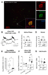
Dser/NMDA membrane and functional responses in a mouse model with β-cell specific GluN1 loss. Ins2-driven Cre-Lox recombination of the Grin1 gene (Rip-cre; Grin1 f/f, βGrin1 KO) was used to achieve β-cell specific loss of D-serine-binding GluN1 protein (and thus all NMDARs) in C57 mice. (A) Immunofluorescent staining of a fixed pancreatic slice (10x, left) and a single islet (20x, right) from a Rip-cre; Grin1 f/+ mouse bred with a CAG-ZsGreen reporter gene showing colocalization of Cre-driven green fluorescence and β-cell insulin signals. (B) Representative recordings of glucose-stimulated (12 mM) membrane voltage oscillations in β-cells from intact βGrin1 KO islets immediately before (black) and ~1 min after D-serine/NMDA (100 μM each, grey) with a comparative quantification of (C) active phase duration and (D) period. (E) The relative effect of the same concentration of D-serine/NMDA on GSIS (22 mM glucose) in WT (Cre-negative; Grin1 f/f or Grin1 f/+) and βGrin1 KO islets and the effect of genotype on islet (F) insulin secretion and (G) insulin content in an independent GSIS experiment. Data in panels C, D were analyzed by 1-sample t-test and in E, G by independent 2-tailed t-test. Data in panel F were analyzed by repeated measures 2-way ANOVA. Not significant (n.s.) p > 0.05, ## p < 0.01 vs. w/in genotype LG or as indicated. * p < 0.05, *** p < 0.001 vs. WT.
Figure 4
Figure 4
In vivo metabolic phenotype of young adult βGrin1 KO mice. WT and βGrin1 KO male and female mice were separately evaluated for random fed (A) body weight, (B) blood glucose, and (C) plasma insulin. (D) Glucose tolerance was assessed in male (circles) and female (triangles) overnight fasted mice and (E) insulin sensitivity in 6-h fasted mice following 0.75 U/kg ip insulin. (F) An in vivo GSIS was also conducted in male mice and (G) β-cell mass was determined by immunofluorescent insulin staining of fixed pancreatic sections from female mice. Statistically, single endpoint data (AC,G), AUC) was assessed by independent 2-tailed t-test and multi-endpoint data (DF) by repeated measures 2-way ANOVA for each sex. ## p < 0.01, ### p < 0.001 vs. w/in genotype fasting level. ITT = Insulin tolerance test. Cre-negative (Grin1 f/f or Grin1 f/+) mice were used as WT controls.
Figure 5
Figure 5
In vivo phenotype of older adult βGrin1 KO mice. Male and female βGrin1 KO and WT mice, aged 7–14 months, were assessed for measures of glucose homeostasis including (A) random fed body weight, (B) blood glucose, and (C) plasma insulin; (D) 12–14-month-old males were also given an IPGTT with AUC shown in the inset. Islet data derived from 7–10-month-old male and female mice from each genotype was combined (diamonds) to show (E) islet insulin secretion by GSIS and (F) total islet insulin content. Statistical analysis was as described for similar data in previous figures, with independent Student t-tests and repeated measures 2-way ANOVA for within sex single and multi-endpoint data, respectively. ### p < 0.001, #### p < 0.0001 vs. within group LG. ** p < 0.01 vs. WT, as indicated. Figure legend defines all subsequent panels until re-defined. Cre-negative (Grin1 f/f or Grin1 f/+) mice were used as WT controls.
Figure 6
Figure 6
Glucose intolerance in young adult pancreatic Grin1 loss mice. A Pdx1-Cre driver was used to partially (pGrin1 Het, grey) or fully (pGrin1 KO, black) knockdown GluN1 protein in all pancreatic tissue; 3–5-month-old mice were phenotyped against Grin1 f/f or f/+ control mice (WT). Male and female random fed mice were evaluated for (A) body weight, (B) blood glucose, and (C) plasma insulin. As previously described, male mice were assessed for (D) glucose tolerance, (E) insulin sensitivity, and (F) in vivo GSIS. Female mice were also tested for (G) glucose tolerance, (H) insulin sensitivity, and (I) in vivo GSIS. Data in all panels were evaluated by repeated measures 2-way ANOVA with Tukey’s (D,G) or Sidak’s (F,I) multiple comparisons. ## p < 0.01, ### p < 0.001 vs. fasting. *, ** p < 0.05, 0.01, vs. WT control.
Figure 7
Figure 7
A hypothetical excitatory model of β-cell NMDAR activity. In canonical β-cell stimulus-secretion coupling, glucose oxidation stimulates the consumption of ADP and production of ATP, which together inhibit K+-effluxing KATP channels, leading to local depolarization and the activation of voltage-sensitive L-type Ca2+ channels, culminating in the Ca2+-triggered exocytosis of insulin granules. When D-serine and NMDA (exogenous) or glutamate (endogenous) are present in the extracellular space during sufficient intracellular depolarization to eject the NMDAR Mg2+ channel block, positive ionic flux through the channel enhances the signal for exocytosis, directly or through a stronger activation/recruitment of Ca2+ channels, resulting in potentiation of insulin secretion. Mechanism of NMDAR inhibition is indicated for 5,7-DCKA and Grin1 KO. TCA = tricarboxylic acid cycle. Ox Phos = Oxidative Phosphorylation.

Similar articles

Cited by

References

    1. Lockridge A.D., Baumann D.C., Akhaphong B., Abrenica A., Miller R.F., Alejandro E.U. Serine racemase is expressed in islets and contributes to the regulation of glucose homeostasis. Islets. 2016;8:195–206. doi: 10.1080/19382014.2016.1260797. - DOI - PMC - PubMed
    1. Wang Y., Nie M., Li W., Ping F., Hu Y., Ma L., Gao J., Liu J. Association of six single nucleotide polymorphisms with gestational diabetes mellitus in a Chinese population. PLoS ONE. 2011;6:e26953. doi: 10.1371/journal.pone.0026953. - DOI - PMC - PubMed
    1. Tsai F.J., Yang C.F., Chen C.C., Chuang L.M., Lu C.H., Chang C.T., Wang T.Y., Chen R.H., Shiu C.F., Liu Y.M., et al. A genome-wide association study identifies susceptibility variants for type 2 diabetes in Han Chinese. PLoS Genet. 2010;6:e1000847. doi: 10.1371/journal.pgen.1000847. - DOI - PMC - PubMed
    1. Dong M., Gong Z.C., Dai X.P., Lei G.H., Lu H.B., Fan L., Qu J., Zhou H.H., Liu Z.Q. Serine racemase rs391300 G/A polymorphism influences the therapeutic efficacy of metformin in Chinese patients with diabetes mellitus type 2. Clin. Exp. Pharmacol. Physiol. 2011;38:824–829. doi: 10.1111/j.1440-1681.2011.05610.x. - DOI - PubMed
    1. Suwandhi L., Hausmann S., Braun A., Gruber T., Heinzmann S.S., Galvez E.J.C., Buck A., Legutko B., Israel A., Feuchtinger A., et al. Chronic d-serine supplementation impairs insulin secretion. Mol. Metab. 2018;16:191–202. doi: 10.1016/j.molmet.2018.07.002. - DOI - PMC - PubMed

Publication types

MeSH terms

LinkOut - more resources