. 2021 Jan 7;13(2):191.
doi: 10.3390/cancers13020191.

A Novel Cyclic Pentadepsipeptide, N-Methylsansalvamide, Suppresses Angiogenic Responses and Exhibits Antitumor Efficacy against Bladder Cancer

Affiliations

A Novel Cyclic Pentadepsipeptide, N-Methylsansalvamide, Suppresses Angiogenic Responses and Exhibits Antitumor Efficacy against Bladder Cancer

Jun-Hui Song et al. Cancers (Basel). .

Abstract

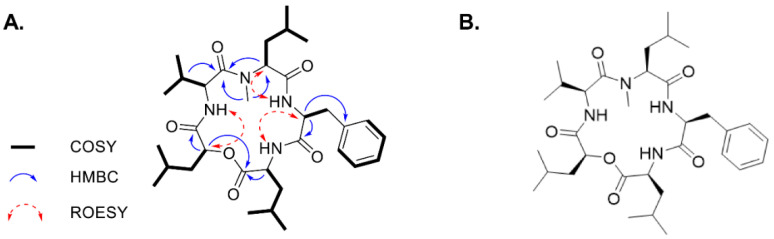
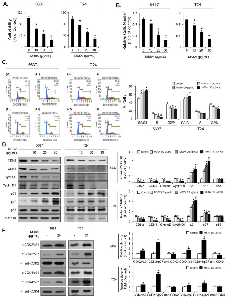
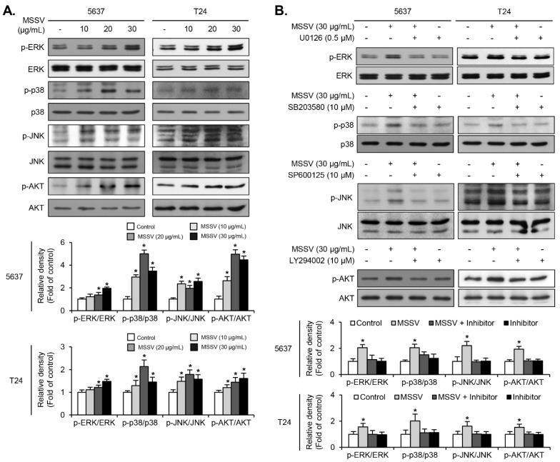
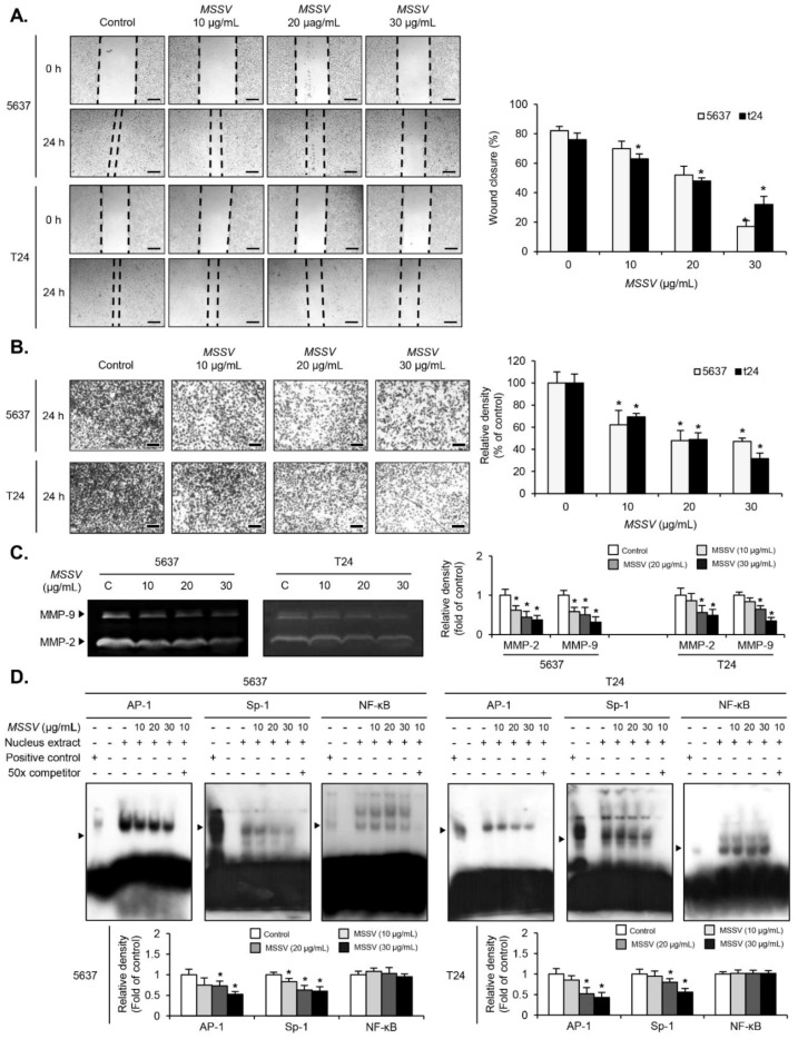
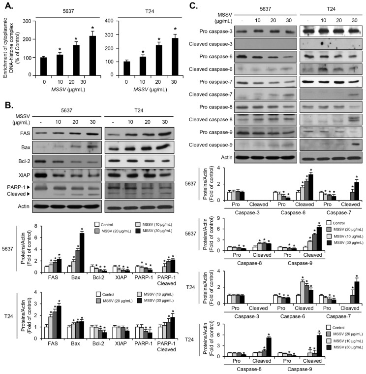
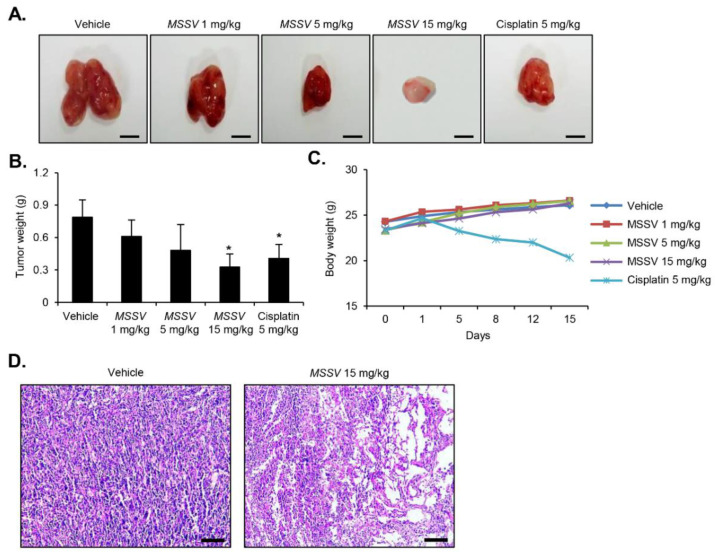
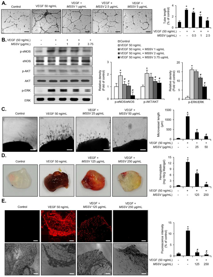
Here, we explored the anti-tumor efficacy of a cyclic pentadepsipeptide, N-methylsansalvamide (MSSV), in bladder cancer. MSSV inhibited the proliferation of both bladder cancer 5637 and T24 cells, which was attributed to the G1-phase cell cycle arrest, apoptosis induction, and alteration of mitogen-activated protein kinases (MAPKs) and protein kinase b (AKT) signaling pathways. Additionally, the treatment of bladder cancer cells with MSSV suppressed migratory and invasive potential via the transcription factor-mediated expression of matrix metalloproteinase 9 (MMP-9). MSSV abrogated vascular endothelial growth factor (VEGF)-induced angiogenic responses in vitro and in vivo. Furthermore, our result showed the potent anti-tumor efficacy of MSSV in a xenograft mouse model implanted with bladder cancer 5637 cells. Finally, acute toxicity test data obtained from blood biochemical test and liver staining indicated that the oral administration of MSSV at 2000 mg/kg caused no adverse cytotoxic effects. Our preclinical data described the potent anti-angiogenic and anti-tumor efficacy of MSSV and showed no signs of acute toxicity, thereby suggesting the putative potential of oral MSSV as a novel anti-tumor agent in bladder cancer treatment.

Keywords: MSSV; anti-angiogenesis; anti-tumor efficacy; bladder cancer; single oral dose of acute toxicity.


Conflict of interest statement

The authors declare no conflict of interest.

Figures

Figure 1
Figure 1
Structure elucidation of N-methylsansalvamide (MSSV). (A) Key correlation spectroscopy (COSY), HMBC, and ROESY correlations of MSSV. (B) Chemical structure of MSSV.
Figure 2
Figure 2
MSSV-mediated inhibition of proliferation of the human bladder cancer cell lines, 5637 and T24, was owing to G1-phase cell cycle arrest. (A) Both cell lines were treated with or without MSSV at the indicated concentrations for 24 h, followed by MTT assays for cell viability. (B) Cells were treated with indicated concentrations of MSSV for 24 h, and cell counting was performed via trypan blue staining. (C) Both cell lines were treated with MSSV for 24 h and analyzed for cell cycle distribution using fluorescence-activated cell sorting (FACS) histograms. The percentage of cells in each cell cycle phase is presented. (D) Cells were exposed to MSSV at the indicated concentrations for 24 h. The expression levels of cyclin D1, cyclin E, CDK2, CDK4, p21WAF1, p27KIP1, p53, and GAPDH were analyzed via immunoblotting. Bar graphs show the relative fold changes in proteins at different MSSV concentrations in comparison with the control. (E) The cell lysates were immunoprecipitated with antibodies recognizing CDK2 and CDK4, followed by immunoblotting with specific antibodies against p21WAF1, p27KIP1, CDK2, and CDK4. Graphs show the relative amount of immunoprecipitated proteins as fold changes in comparison with the control. For the bar graphs, values were presented as the mean ± SD of three independent experiments; * p < 0.05, compared with the control group. Uncropped Western Blot Images in Figures S3 and S4.
Figure 3
Figure 3
Signaling pathways mediating the anti-proliferative effect of MSSV in bladder cancer cells. (A) Both cells were treated with MSSV at the indicated concentrations for 24 h. The cell lysates were subjected to immunoblot analysis with phosphorylated and total forms of MAPKs (ERK1/2, JNK1/2, and p38) and AKT; Results were expressed as fold changes in comparison with the control cell lysates. (B) Both cells were pretreated with biochemical inhibitors of U0126 (0.5 μM), SB203580 (10 μM), SP600125 (10 μM), and LY 294002 (10 μM) for 40 min. MSSV was then added for another 24 h, followed by immunoblot analysis. The ratio of phosphorylated to non-phosphorylated form was measured and expressed as fold changes in comparison with the ratio associated with MSSV treatment. For the bar graphs, values are presented as the mean ± SD of three independent experiments; * p < 0.05, compared with the control group. Uncropped Western Blot Images in Figures S5 and S6.
Figure 4
Figure 4
MSSV represses the migration and invasion of bladder cancer cells by suppressing transcription factor-mediated MMP-9 activity. Both 5637 and T24 cells were pretreated with mitomycin C, followed by incubation with MSSV for 24 h. (A) Cellular images of migratory recovery rate of 5637 and T24 cells photographed under an inverted microscope (scale bars = 200 µm). Bar graphs represent the relative fold changes in migration distances as compared with the control. (B) Treatment with MSSV inhibited the invasion ability of both bladder cancer cells (scale bars = 200 µm). Both cells were added onto the upper chamber and incubated with the indicated concentrations of MSSV for 24 h. Cells invading the lower surface of the membrane were detected by crystal violet staining. In the bar graphs, the amount of invading cells was assessed as the fold change compared with the control. (C) Zymographic assay was performed to determine MMP-9 expression in cells isolated from the cultured medium. Proteolytic activity of each MMP-9 was detected as a fold change compared with the control. (D) Nuclear extracts were subjected to EMSA assay to test the binding activities of activator protein 1 (AP-1), specificity protein 1 (Sp-1), and nuclear factor kappa-light-chain-enhancer of activated B cells (NF-κB) using radiolabeled oligonucleotide probes. Unlabeled AP-1, Sp-1, and NF-κB oligonucleotides were used as competitors. For the bar graphs, values are expressed as the mean ± SD of three independent experiments; * p < 0.05, compared with the control group.
Figure 5
Figure 5
Effect of MSSV on apoptosis signaling in both bladder cancer cells. Both cells were exposed to MSSV at the indicated concentrations for 24 h. (A) Detection of apoptosis via measurement of cytoplasmic DNA-histone complex. After the treatment of cells with MSSV, supernatants containing cytoplasmic histone-associated DNA fragments were determined using ELISA assay. (B) Whole-cell lysates were subjected to immunoblot analysis with anti-FAS, anti-Bcl-2, anti-Bax, anti-XIAP, anti-PARP-1, and anti-Actin. (C) Expression levels of pro and cleaved forms of cascase-3, cascase-6, cascase-7, cascase-8, and cascase-9 were analyzed via immunoblotting. Actin was used as the internal control. Bar graphs show relative fold changes in the protein levels at different concentrations of MSSV compared with the control. For the bar graphs, values are presented as the mean ± SD of three independent experiments; * p < 0.05, compared with the control group. Uncropped Western Blot Images in Figures S7 and S8.
Figure 6
Figure 6
Antitumor efficacy of MSSV in bladder cancer 5637 xenograft mice models. (A) MSSV administration via oral gavage twice a day caused the inhibition of tumor growth in bladder cancer-xenografted mouse model (scale bars = 50 mm). (B,C) Tumor weight and body weight are represented in the bar graph. All data are represented as the means ± SE from three independent experiments. * p < 0.05, compared with control. (D) Tumor tissues were subjected to H&E staining for the detection of tumors (scale bars = 50 µm).
Figure 7
Figure 7
Effect of MSSV in VEGF-induced angiogenic responses in vitro, ex vivo, and in vivo. (A) HUVECs were treated with VEGF (50 ng/mL) for 1 h, followed by the indicated concentrations of MSSV for 24 h. The HUVEC tube formation assay was accomplished using Matrigel (scale bars = 200 µm). (B) Immunoblot analysis for antibodies specific for phospho-ERK1/2, ERK1/2, phospho-AKT, AKT, phospho-eNOS, and eNOS. (C) Blood neo-vessel sprouting in the mouse aorta after MSSV treatment in the presence of VEGF (50 ng/mL) was evaluated using aortic ring assay (scale bars = 200 µm). Bar graphs show the relative fold changes in the number of emerging neo-vessel associated with MSSV + VEGF treatment in comparison with that associated with VEGF treatment alone. (D) Blood vessel formation was performed using a Matrigel plug in an in vivo experiment. Blood vessel formation was assessed by measuring hemoglobin content in Matrigel. Bar graphs show the relative fold changes in the hemoglobin content associated with MSSV + VEGF treatment in comparison with that associated with VEGF treatment alone. (E) Analysis of vessel formation in Matrigel plug was performed by CD31 immunostaining (scale bars = 100 µm). Bar graphs show the relative fold changes in the density of CD31-positive vessels during MSSV + VEGF treatment as compared with that during VEGF treatment alone. All data are represented as the mean ± SE from three independent experiments. * p < 0.05 compared with control and # p < 0.05 compared with VEGF treatment. Uncropped Western Blot Images in Figure S9.

Similar articles

Cited by

References

    1. Metts M.C., Metts J.C., Milito S.J., Thomas C.R. Bladder Cancer: A Review of Diagnosis and Management. J. Natl. Med. Assoc. 2000;92:285–294. - PMC - PubMed
    1. Shen Z., Shen T., Wientjes M.G., O’Donnell M.A., Au J.L. Intravesical Treatments of Bladder Cancer: Review. Pharm. Res. 2008;25:1500–1510. doi: 10.1007/s11095-008-9566-7. - DOI - PMC - PubMed
    1. Black P.C., Dinney C.P.N. Bladder Cancer Angiogenesis and Metastasis—Translation from Murine Model to Clinical Trial. Cancer Metastasis Rev. 2007;26:623–634. doi: 10.1007/s10555-007-9084-9. - DOI - PubMed
    1. Zachos I., Konstantinopoulos P.A., Tzortzis V., Gravas S., Karatzas A., Karamouzis M.V., Melekos M., Papavassiliou A.G. Systemic Therapy of Metastatic Bladder Cancer in the Molecular Era: Current Status and Future Promise. Expert Opin. Investig. Drugs. 2010;19:875–887. doi: 10.1517/13543784.2010.496450. - DOI - PubMed
    1. Yun S.J., Moon S., Kim W. Investigational Cell Cycle Inhibitors in Clinical Trials for Bladder Cancer. Expert Opin. Investig. Drugs. 2013;22:369–377. doi: 10.1517/13543784.2013.751097. - DOI - PubMed

LinkOut - more resources