

Vessel wall MRI in ruptured cranial dural arteriovenous fistulas
- PMID: 33430655
- PMCID: PMC8580544
- DOI: 10.1177/1591019920988205
Vessel wall MRI in ruptured cranial dural arteriovenous fistulas
Abstract
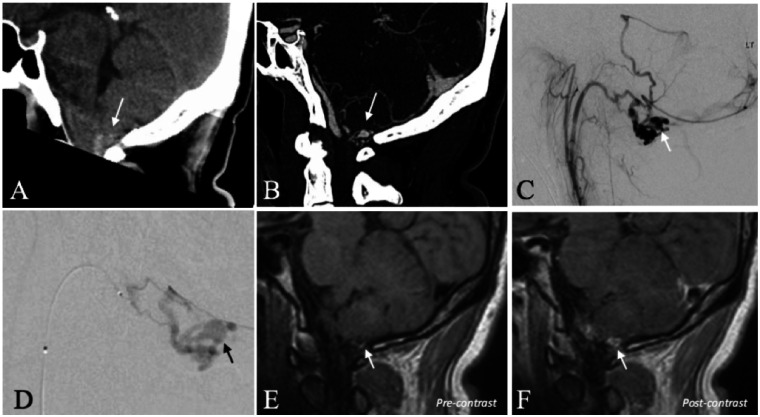
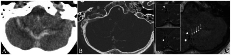
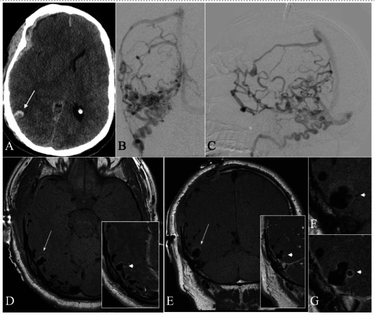
Intracranial high-resolution vessel wall MRI (VW-MRI) is an imaging paradigm that is useful in site-of-rupture identification in patients presenting with spontaneous subarachnoid hemorrhage and multiple intracranial aneurysms. Only a handful of case reports describe its potential utility in the evaluation of more complex brain vascular malformations. We report for the first time three patients with ruptured cranial dural arteriovenous fistulas (dAVFs) that were evaluated with high-resolution VW-MRI. The presumed site-of-rupture was identified based on contiguity of a venous ectasia with adjacent blood products and thick, concentric wall enhancement. This preliminary experience suggests a role for high-resolution VW-MRI in the evaluation of ruptured cranial dAVFs, in particular, site-of-rupture identification. It also supports an emerging hypothesis that all spontaneously ruptured, macrovascular lesions demonstrate avid vessel wall enhancement.
Keywords: Dural arteriovenous fistula; magnetic resonance; vessel wall MRI.
Conflict of interest statement
Figures
Similar articles
-
Dural arteriovenous fistula of the lumbar spine presenting with subarachnoid hemorrhage. Case report and review of the literature.J Neurosurg. 2004 Apr;100(4 Suppl Spine):385-91. doi: 10.3171/spi.2004.100.4.0385. J Neurosurg. 2004. PMID: 15070151
-
Vessel wall enhancement of intracranial aneurysms: fact or artifact?Neurosurg Focus. 2019 Jul 1;47(1):E18. doi: 10.3171/2019.4.FOCUS19236. Neurosurg Focus. 2019. PMID: 31261122
-
High-Resolution Vessel Wall Magnetic Resonance Imaging in Angiogram-Negative Non-Perimesencephalic Subarachnoid Hemorrhage.Clin Neuroradiol. 2017 Jun;27(2):175-183. doi: 10.1007/s00062-015-0484-x. Epub 2015 Nov 25. Clin Neuroradiol. 2017. PMID: 26608742
-
Pial-Dural Intracranial Arteriovenous Fistula with Flow-Associated Aneurysmal Rupture-Case Report with Review of Literature and Proposal on the Mechanism of Hemorrhage and Treatment Options.World Neurosurg. 2017 Sep;105:1040.e15-1040.e19. doi: 10.1016/j.wneu.2017.06.152. Epub 2017 Jul 1. World Neurosurg. 2017. PMID: 28676463 Review.
-
Midterm Follow-Up of 20 Consecutive Patients with Nonaneurysmal Subarachnoid Hemorrhage of Unknown Origin in a Single-Center: Two Cases of De Novo Development of Dural Arteriovenous Fistula.J Stroke Cerebrovasc Dis. 2017 Dec;26(12):2788-2792. doi: 10.1016/j.jstrokecerebrovasdis.2017.06.061. Epub 2017 Aug 10. J Stroke Cerebrovasc Dis. 2017. PMID: 28802521 Review.
Cited by
-
Estimation of the rupture point of the craniovertebral junction intradural arteriovenous fistula with vessel wall magnetic resonance image and its pathological findings: A case report.Surg Neurol Int. 2024 May 3;15:149. doi: 10.25259/SNI_163_2024. eCollection 2024. Surg Neurol Int. 2024. PMID: 38742004 Free PMC article.
-
Overview of multimodal MRI of intracranial Dural arteriovenous fistulas.J Interv Med. 2022 Nov 28;5(4):173-179. doi: 10.1016/j.jimed.2022.04.004. eCollection 2022 Nov. J Interv Med. 2022. PMID: 36532312 Free PMC article.
-
Vasa vasorum: The role in intracranial physiology and pathophysiology.Surg Neurol Int. 2024 Jun 7;15:188. doi: 10.25259/SNI_214_2024. eCollection 2024. Surg Neurol Int. 2024. PMID: 38974550 Free PMC article. Review.
References
-
- Davies MA, TerBrugge K, Willinsky R, et al.. The validity of classification for the clinical presentation of intracranial dural arteriovenous fistulas. J Neurosurg 1996; 85: 830–837. - PubMed
-
- Davies MA, Saleh J, Ter Brugge K, et al.. The natural history and management of intracranial dural arteriovenous fistulae. Part 1: benign lesions. Interv Neuroradiol 1997; 3: 295–302. - PubMed
-
- Matouk CC, Mandell DM, Günel M, et al.. Vessel wall magnetic resonance imaging identifies the site of rupture in patients with multiple intracranial aneurysms: proof of principle. Neurosurgery 2013; 72: 492–496. - PubMed
-
- Omodaka S, Endo H, Niizuma K, et al.. Circumferential wall enhancement on magnetic resonance imaging is useful to identify rupture site in patients with multiple cerebral aneurysms. Neurosurgery 2018; 82: 638–644. - PubMed
-
- Texakalidis P, Hilditch CA, Lehman V, et al.. Vessel wall imaging of intracranial aneurysms: systematic review and meta-analysis. World Neurosurg 2018; 117: 453–458.e1. - PubMed
MeSH terms
Grants and funding
LinkOut - more resources
Full Text Sources
Other Literature Sources
Medical
