. 2021 Jan 10;10(1):bio057539.
doi: 10.1242/bio.057539.

Bisphenol A promotes stress granule assembly and modulates the integrated stress response

Affiliations

Bisphenol A promotes stress granule assembly and modulates the integrated stress response

Marta M Fay et al. Biol Open. .

Abstract

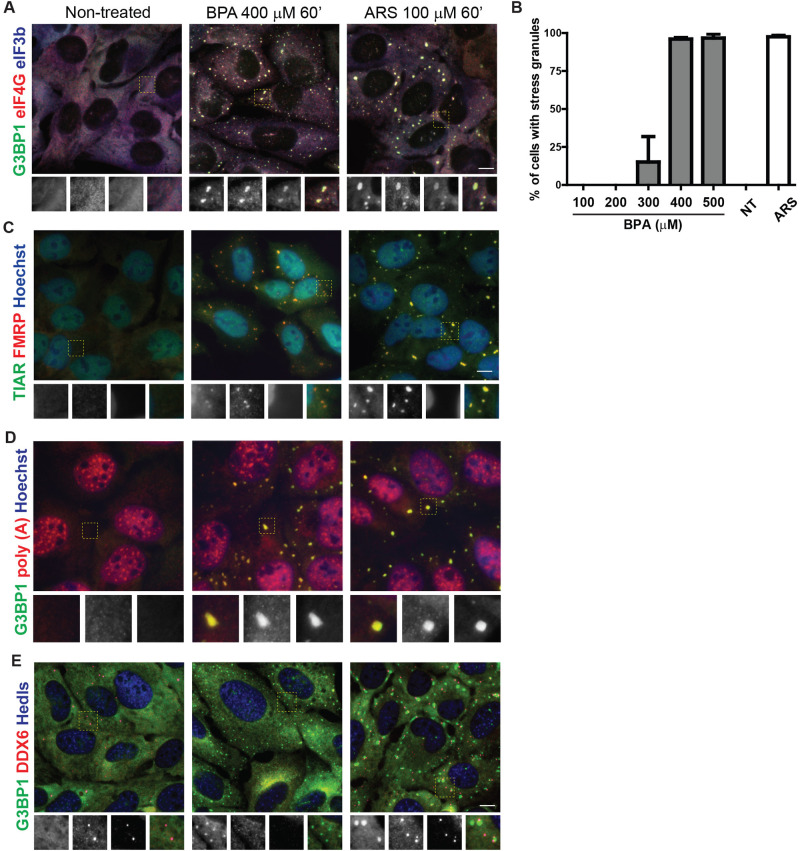
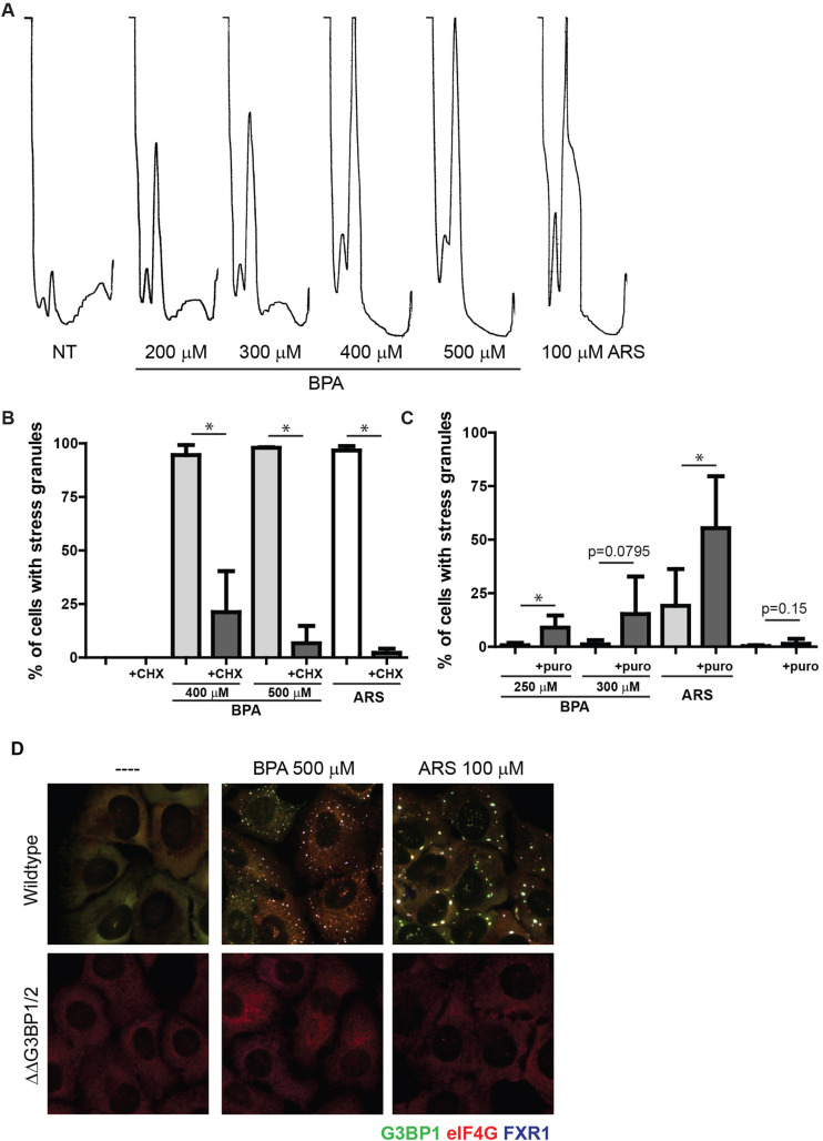
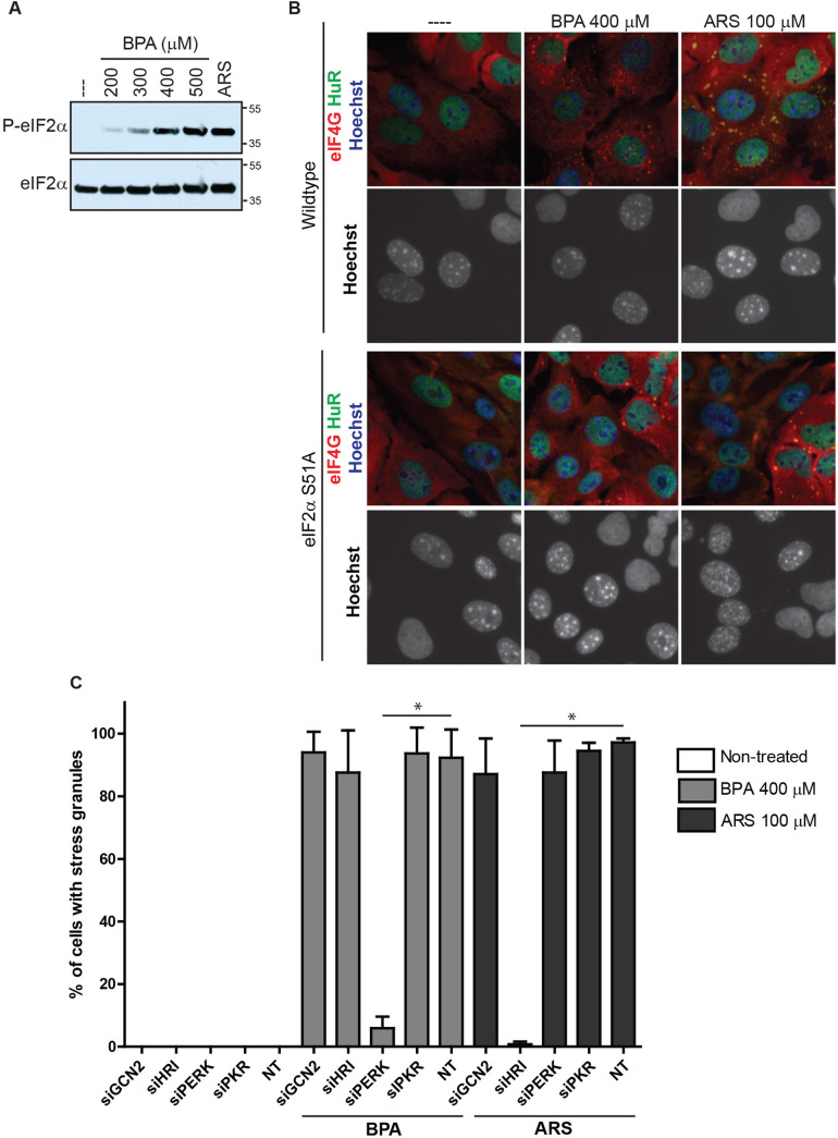
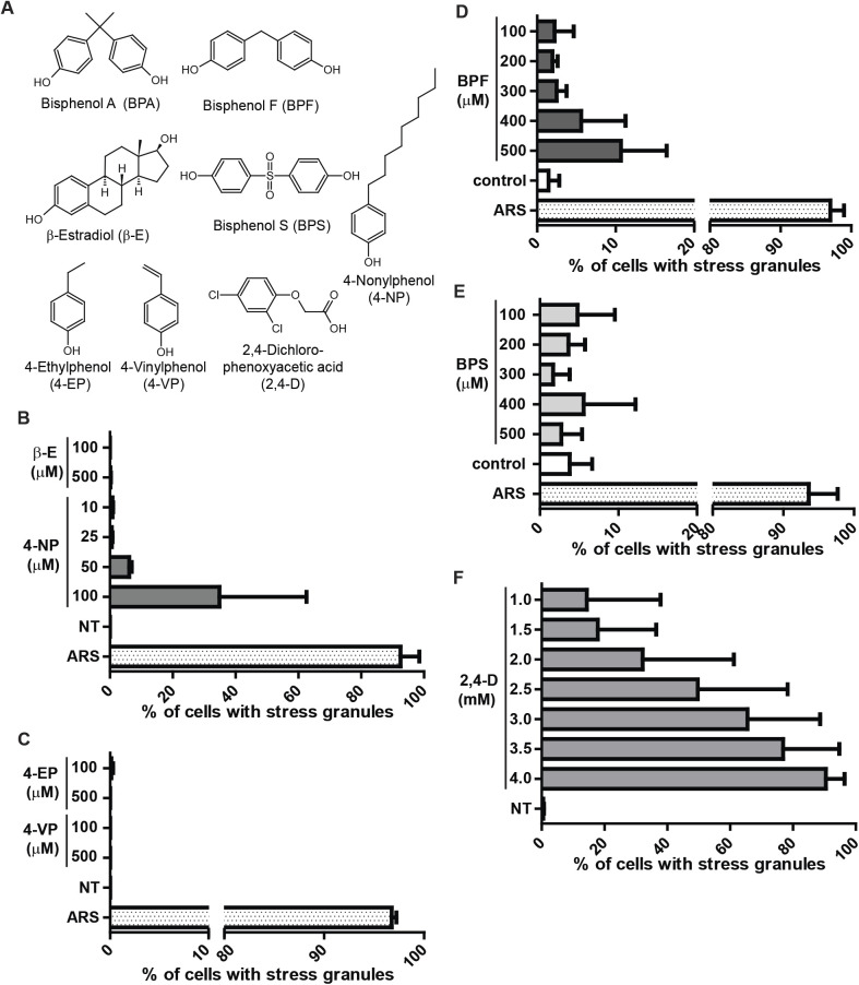
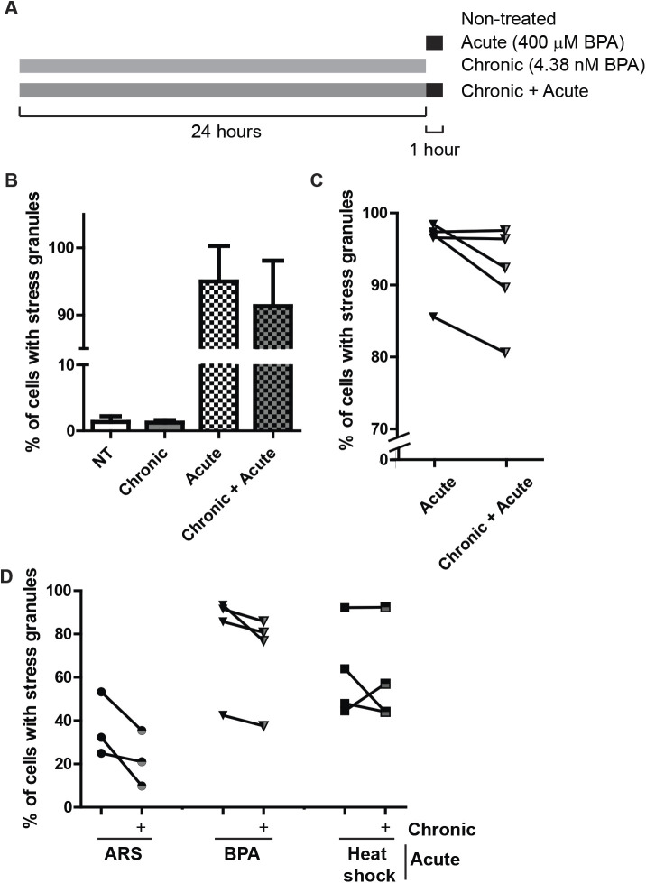
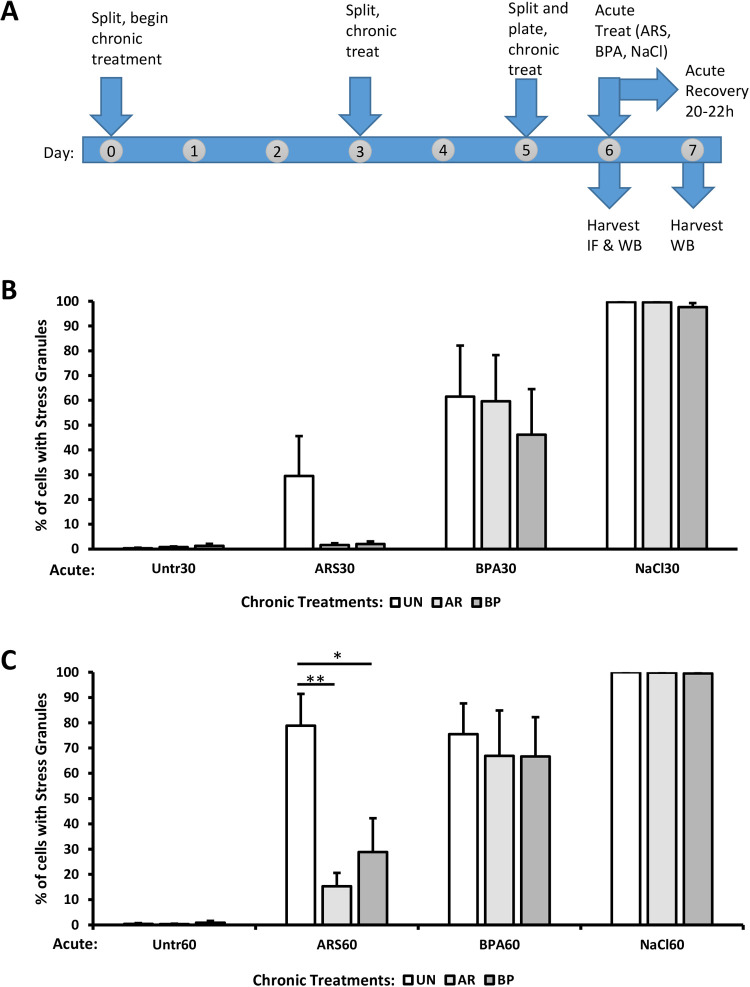
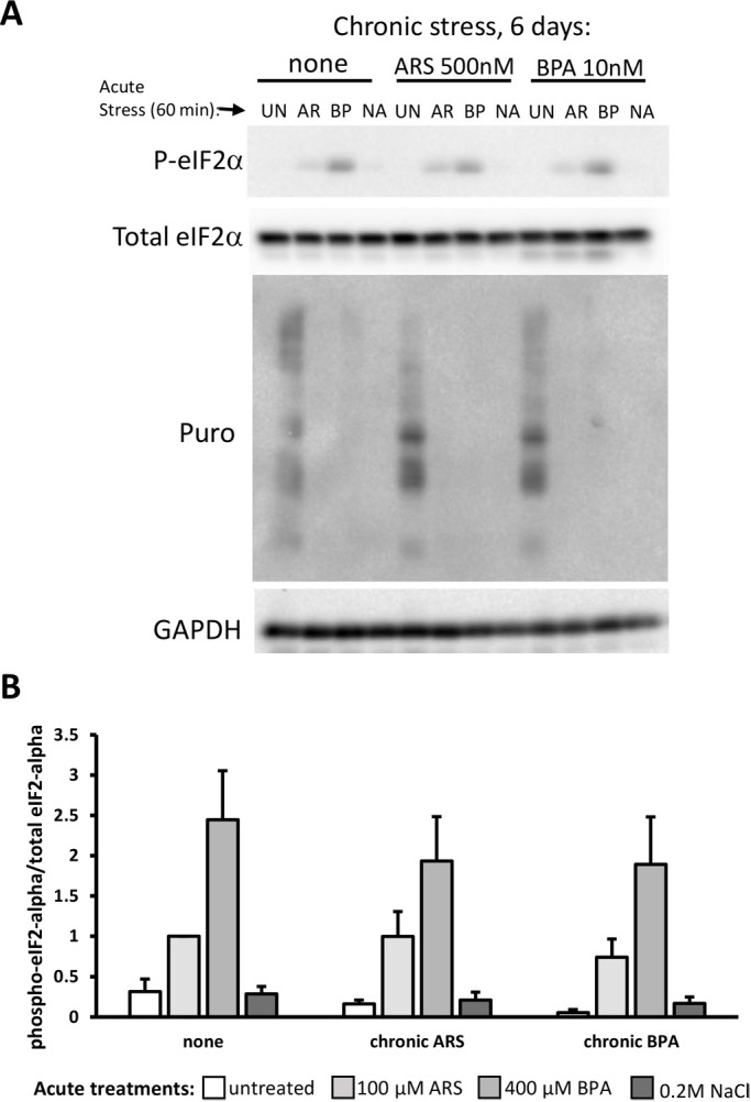
Bisphenol-A (BPA) is a ubiquitous precursor of polycarbonate plastics that is found in the blood and serum of >92% of Americans. While BPA has been well documented to act as a weak estrogen receptor (ER) agonist, its effects on cellular stress are unclear. Here, we demonstrate that high-dose BPA causes stress granules (SGs) in human cells. A common estrogen derivative, β-estradiol, does not trigger SGs, indicating the mechanism of SG induction is not via the ER pathway. We also tested other structurally related environmental contaminants including the common BPA substitutes BPS and BPF, the industrial chemical 4-nonylphenol (4-NP) and structurally related compounds 4-EP and 4-VP, as well as the pesticide 2,4-dichlorophenoxyacetic acid (2,4-D). The variable results from these related compounds suggest that structural homology is not a reliable predictor of the capacity of a compound to cause SGs. Also, we demonstrate that BPA acts primarily through the PERK pathway to generate canonical SGs. Finally, we show that chronic exposure to a low physiologically relevant dose of BPA suppresses SG assembly upon subsequent acute stress. Interestingly, this SG inhibition does not affect phosphorylation of eIF2α or translation inhibition, thus uncoupling the physical assembly of SGs from translational control. Our work identifies additional effects of BPA beyond endocrine disruption that may have consequences for human health.

Keywords: Bisphenol-A; Integrated stress response; Stress granules; Translational control.


Conflict of interest statement

Competing interestsThe authors declare no competing or financial interests.

Figures

Fig. 1.
Fig. 1.
BPA promotes SG formation. (A) Immunofluorescence of U2OS cells untreated (left) or treated with BPA (400 µM) (center) or arsenite (ARS, 100 µM) for 1 h (right) detecting G3BP1 (green), eIF4G (red), eIF3b (blue). Boxed area is shown zoomed at 1.8X of original and individual channels are shown as black and white in order of green, red, blue then merged as a RGB image. (B) U2OS cells were treated with the indicated range of BPA concentrations or 100 µM ARS for 1 h, or left untreated (NT) then assessed for SG formation by immunofluorescence. Quantification of SGs was determined by the total number of cells containing two or more G3BP1 positive foci over the total number of cells. (C) Same as A, but detecting TIAR (green), FMRP (red), and Hoechst (blue). (D) U2OS cells treated as indicated in B then fluorescence in situ hybridization to detect poly (A) RNAs (red) followed by immunofluorescence to detect G3BP1 (green) and counterstained with Hoechst (blue). (E) Same as B but detecting G3BP1 (green), DDX6 (red), and Hedls (blue). Scale bar indicates 10 microns.
Fig. 2.
Fig. 2.
BPA induced SGs requires polysome disassembly and G3BP1/2 mediated condensation. (A) Polysome profiles of U2OS cells treated with indicated concentrations of BPA or ARS for 60 min. Profiles were generated using OD 254 nm. (B) U2OS cells were treated with BPA or ARS (100 µM) for 60 min. Where indicated, 50 µg/ml of cycloheximide was added for the final 30 min. Immunofluorescence was performed and used to quantify the percentage of cells with SGs. Scale bar indicates 10 microns. (C) U2OS cells were treated with indicated concentrations of BPA or ARS (50 µM) for 60 min, and co-treated with 20 µg/mL puromycin where indicated. Immunofluorescence analysis was used to quantify the percentage of cells with SGs. (D) G3BP1 (green), eIF4G (red), and FXR1 (blue) in wild-type U2OS cells (top panel), or U2OS cells lacking G3BP1/2 (ΔΔG3BP1/2; bottom panel) treated with indicated concentration of BPA or ARS for 60 min or left untreated. (B,C) Graphs represent mean with standard deviation; n≥3; * 0.05≥P.
Fig. 3.
Fig. 3.
BPA-induced SG assembly requires PERK mediated phosphorylation of eIF2α. (A) Immunoblot detecting levels of phosphorylated eIF2α (top blot, labeled P-eIF2α) and total eIF2α (bottom blot) in U2OS cells treated as indicated with BPA, ARS (100 µM) for 1 h, or left untreated (indicated as ---). (B) Immunofluorescence detecting HuR (green), eIF4G (red), and Hoechst/DNA (blue) in wild-type MEFs co-cultured with U2OS cells (top panels), or MEFs with eIF2α-S51A point mutation co-cultured with U2OS cells (bottom panels), treated as indicated with BPA or ARS for 1 h or left untreated. MEFs exhibit punctate nuclear DNA. Scale bar indicates 10 microns. (C) Quantification of SGs in U2OS cells following siRNA-mediated knockdown of GCN2, HRI, PERK, and PKR, prior to 1 h BPA or ARS treatment, or no treatment. Graph represents mean with standard deviation; n≥3; * 0.05≥P.
Fig. 4.
Fig. 4.
The BPA replacement BPF, the industrial chemical 4-NP, and the pesticide 2,4-D cause SG assembly. (A) Molecular structures of chemicals used. Structures generated using ChemSketch. (B) Quantification of SGs assessed by immunofluorescence in U2OS cells treated with indicated concentrations of β-estradiol (β-E), 4-nonylphenol (4-NP), or ARS for 1 h or left untreated (NT). (C) Same as B, but treated with 4-ethylphenol (4-EP), or 4-vinylphenol (4-VP). (D–F) Quantification of SGs assessed by direct fluorescence GFP-G3BP1 in stably expressing U2OS cells treated with indicated concentrations of BPF, BPS, 2,4-dichlorophenoxyacetic acid (2,4-D), or ARS for 1 h or mock treated (control). (B–F): Graphs represent means with standard deviation; n≥3; * 0.05≥P.
Fig. 5.
Fig. 5.
Chronic BPA exposure using physiologically relevant doses partially inhibits SGs assembly. (A) Schematic indicating experiment performed to generate data in B and C. (B) Quantification of SGs assesed by direct fluorescence of GFP-G3BP1 in stably expressing U2OS cells treated with BPA by acute (400 µM, 60 min), chronic (4.38 nM, 24 h) or both chronic and acute treatment. (C) Same experiment (and data) as B showing how experimental samples pair as indicated by the line. Error bars in B and C represent +/- one standard deviation. (D) Quantification of SGs detected by immunofluorescence in U2OS long term, chronically BPA-treated cells (5 nM, 1 month; half grey half black shapes) or cells left untreated (solid black shapes) then treated with 75 µM ARS or 350 µM BPA for 60 min or heat shocked at 42°C for 25 min. Lines between chronically BPA-treated and untreated samples indicate experimentally paired samples. n=3–5 replicates per condition, as indicated by paired data in C and D.
Fig. 6.
Fig. 6.
Chronic BPA or ARS suppresses eIF2α-dependent SG formation. (A) Schematic of the protocol for experiments in Figs 6B,C and 7. Cells were not treated (UN, white bars) or chronically treated for 6 days with 500 nM arsenite (AR, light grey bars), or 10 nM BPA (BP, dark grey bars). After chronic treatment protocol, acute stresses (100 μM arsenite, 400 μM BPA, or 0.2 M sodium chloride) were applied for 30 min (B) or 60 min (C), followed by fixation or processing for western blotting. An additional experimental replicate was treated for 60 min with acute stress and then recovered for 20-22 h before processing for western blotting or Coomassie stain (see Fig. S4). n=4; error bars are ±s.e.m.; *P<0.05, **P<0.01 by one-way ANOVA within acute treatment groups.
Fig. 7.
Fig. 7.
Chronic BPA or ARS exposure does not affect eIF2α phosphorylation or translational arrest. (A,B) Western blotting (A) of U2OS cells treated with chronic stress for 6 days (none, 500 nM arsenite, 10 nM BPA), followed by acute stress treatments with 100 μM arsenite (AR, light grey bars), 400 μM BPA (BP, medium grey bars) or 0.2 M sodium chloride (NA, dark grey bars) or untreated (UN, white bars). Puromycin (5 µg/ml) was added to the last 5 min of the 60-min acute treatment before harvesting cells for western blotting with the antibodies indicated. (B) Quantification of the phospho-eIF2α blot relative to total eIF2α. No significant difference in the pattern of responses among chronic treatments was observed by two-way ANOVA; n=3; error bars are ±s.e.m..

References

    1. Anderson P. and Kedersha N. (2009). Stress granules. Curr. Biol. 19, R397-R398. 10.1016/j.cub.2009.03.013 - DOI - PubMed
    1. Anderson P., Kedersha N. and Ivanov P. (2015). Stress granules, P-bodies and cancer. Biochim. Biophys. Acta 1849, 861-870. 10.1016/j.bbagrm.2014.11.009 - DOI - PMC - PubMed
    1. Apau J., Acheampong A. and Adua E (2018). Exposure to Bisphenol A, Bisphenol F, and Bisphenol S can result in obesity in human body. Cogent Chem. 4, 1-7. 10.1080/23312009.2018.1506601 - DOI
    1. Arimoto K., Fukuda H., Imajoh-Ohmi S., Saito H. and Takekawa M. (2008). Formation of stress granules inhibits apoptosis by suppressing stress-responsive MAPK pathways. Nat. Cell Biol. 10, 1324-1332. 10.1038/ncb1791 - DOI - PubMed
    1. Asimakopoulos A. G. and Thomaidis N. S. (2015). Bisphenol A, 4-t-octylphenol, and 4-nonylphenol determination in serum by hybrid solid phase extraction-precipitation technology technique tailored to liquid chromatography-tandem mass spectrometry. J. Chromatogr. B Analyt. Technol. Biomed. Life Sci. 986-987, 85-93. 10.1016/j.jchromb.2015.02.009 - DOI - PubMed

Publication types