Case Reports
. 2021 Jan 11;14(1):e239199.
doi: 10.1136/bcr-2020-239199.

Unusual presentation of mucormycosis mimicking a localised sino-orbital pathology

Affiliations
Case Reports

Unusual presentation of mucormycosis mimicking a localised sino-orbital pathology

Anuj Dadhich et al. BMJ Case Rep. .

Abstract

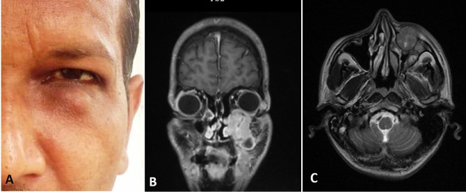
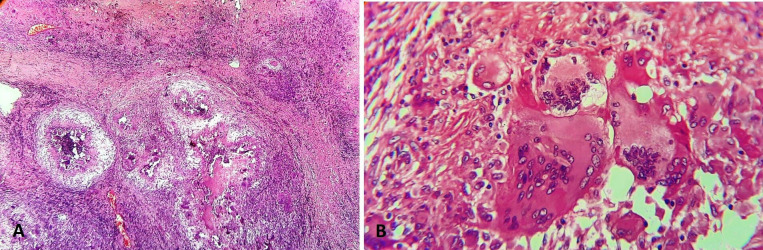
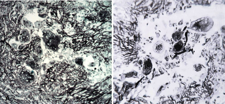
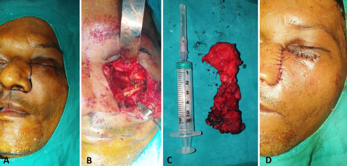
Mucormycosis is an aggressive and deadly fungal infection, which is invariably associated with an immunocompromised patient. Mucormycosis in the head and neck region presents as skeletal necrosis, with or without soft tissue involvement. Early identification and treatment with combination of surgical debridement and parenteral antifungal therapy is critical for a favourable outcome. This paper reports an unusual presentation of mucormycosis, mimicking a localised sino-orbital pathology involving the infraorbital subcutaneous tissue and the maxillary sinus, in a 35 years old immunocompetent man. Despite aggressive antifungal therapy and surgical management, the course of disease was fatal, reiterating the high mortality associated with mucormycosis.

Keywords: oral and maxillofacial surgery; otolaryngology / ENT.


Conflict of interest statement

Competing interests: None declared.

Figures

Figure 1
Figure 1
(A) Preoperative clinical photograph showing localised subcutaneous swelling in left infraorbital area and MRI (B) coronal and (C) axial sections showing well-defined lesion involving the anterior wall of the left maxillary sinus.
Figure 2
Figure 2
H&E stained sections showing multiple caseating granulomas with numerous giant cells (10x) (A) and foreign body type of giant cells with fungal elements (40x) (B).
Figure 3
Figure 3
Gomori methamine silver stained photomicrograph demonstrating fungal elements within the necrotic material and giant cells, seen in 10x (A) and 40x magnification (B).
Figure 4
Figure 4
Intraoperative images showing (A) incision marking, (B) surgical site, (C) excised lesion and (D) closure.

References

    1. Bhansali A, Bhadada S, Sharma A, et al. . Presentation and outcome of rhino-orbital-cerebral mucormycosis in patients with diabetes. Postgrad Med J 2004;80:670–4. 10.1136/pgmj.2003.016030 - DOI - PMC - PubMed
    1. Shamim MS, Siddiqui AA, Enam SA, et al. . Craniocerebral aspergillosis in immunocompetent hosts: surgical perspective. Neurol India 2007;55:274–81. 10.4103/0028-3886.35689 - DOI - PubMed
    1. Roden MM, Zaoutis TE, Buchanan WL, et al. . Epidemiology and outcome of zygomycosis: a review of 929 reported cases. Clin Infect Dis 2005;41:634–53. 10.1086/432579 - DOI - PubMed
    1. Nilesh K, Vande AV. Mucormycosis of maxilla following tooth extraction in immunocompetent patients: reports and review. J Clin Exp Dent 2018;10:e300–5. - PMC - PubMed
    1. Kirszrot J, Rubin PAD. Invasive fungal infections of the orbit. Int Ophthalmol Clin 2007;47:117–32. 10.1097/IIO.0b013e31803776db - DOI - PubMed

Publication types