Prognostic potential of cardiac structural and functional parameters and N-terminal propeptide of type III procollagen in predicting cardiac fibrosis one year after myocardial infarction with preserved left ventricular ejection fraction
- PMID: 33431713
- PMCID: PMC7835023
- DOI: 10.18632/aging.202495
Prognostic potential of cardiac structural and functional parameters and N-terminal propeptide of type III procollagen in predicting cardiac fibrosis one year after myocardial infarction with preserved left ventricular ejection fraction
Abstract
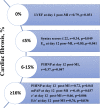
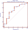
The aim of the study were to evaluate the prognostic potential of serum level of N-terminal propeptide procollagen type III (PIIINP) and heart parameters for predicting heart cardiac fibrosis 1 year after ST-segment elevation myocardial infarction (STEMI) with preserved left ventricular ejection fraction (LVEF). 68 patients with STEMI and preserved LVEF with acute heart failure of the I-III degree according to the Killip classification were examined. Echocardiography was performed and PIIINP levels were measured on days 1 and 12, as well as 1 year after STEMI. A year after STEMI, was performed contrast magnetic resonance imaging and patients were assigned into four groups depending on the severity of cardiac fibrosis: cardiac fibrosis 0% (n=49, 57% of 86 patients); ≤5% (n=18, 20.9%); 6-15% (n=10, 11.6%); ≥16% (n=9, 10.5%). Direct correlations between the severity of cardiac fibrosis, PIIINP level and indicators of diastolic function were established. The risk of cardiac fibrosis increases at the level of PIIINP ≥381.4 ng / ml on the 12th day after STEMI with preserved LVEF (p=0.048). Thus, measuring the level of PIIINP in the inpatient period can allow timely identification of patients with a high risk of cardiac fibrosis 1 year after STEMI with preserved LVEF.
Keywords: cardiofibrosis; diastolic dysfunction; heart failure; myocardial infarction.
Conflict of interest statement
Figures


References
-
- Ambale-Venkatesh B, Liu CY, Liu YC, Donekal S, Ohyama Y, Sharma RK, Wu CO, Post WS, Hundley GW, Bluemke DA, Lima JA. Association of myocardial fibrosis and cardiovascular events: the multi-ethnic study of atherosclerosis. Eur Heart J Cardiovasc Imaging. 2019; 20:168–76. 10.1093/ehjci/jey140 - DOI - PMC - PubMed
-
- Benjamin EJ, Virani SS, Callaway CW, Chamberlain AM, Chang AR, Cheng S, Chiuve SE, Cushman M, Delling FN, Deo R, de Ferranti SD, Ferguson JF, Fornage M, et al., and American Heart Association Council on Epidemiology and Prevention Statistics Committee and Stroke Statistics Subcommittee. Heart disease and stroke statistics-2018 update: a report from the American Heart Association. Circulation. 2018; 137:e67–492. 10.1161/CIR.0000000000000558 - DOI - PubMed
-
- Gandhi PU, Gaggin HK, Redfield MM, Chen HH, Stevens SR, Anstrom KJ, Semigran MJ, Liu P, Januzzi JL Jr. Insulin-like growth factor-binding protein-7 as a biomarker of diastolic dysfunction and functional capacity in heart failure with preserved ejection fraction: results from the RELAX trial. JACC Heart Fail. 2016; 4:860–69. 10.1016/j.jchf.2016.08.002 - DOI - PMC - PubMed
Publication types
MeSH terms
Substances
LinkOut - more resources
Full Text Sources
Other Literature Sources
Medical

