. 2021 Jan 11;12(1):216.
doi: 10.1038/s41467-020-20458-9.

Isolation of MERS-related coronavirus from lesser bamboo bats that uses DPP4 and infects human-DPP4-transgenic mice

Affiliations

Isolation of MERS-related coronavirus from lesser bamboo bats that uses DPP4 and infects human-DPP4-transgenic mice

Susanna K P Lau et al. Nat Commun. .

Abstract

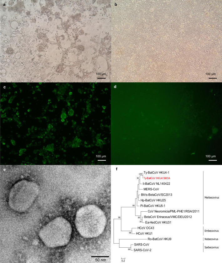
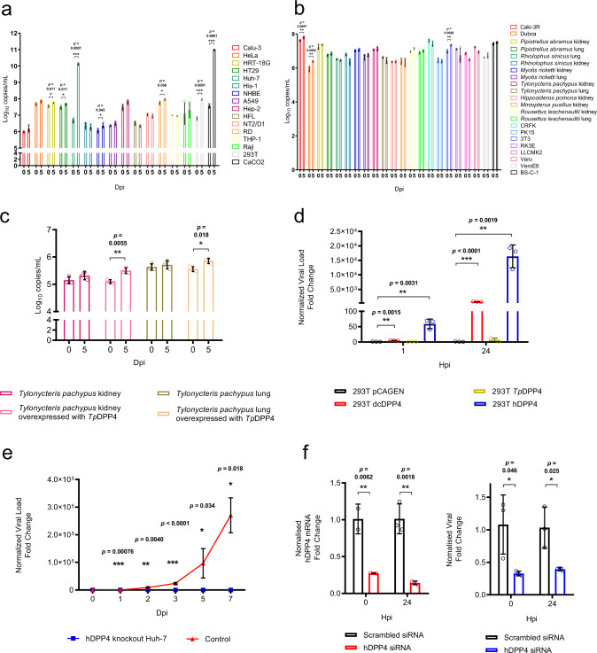
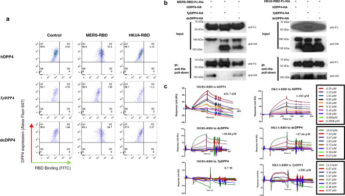
While a number of human coronaviruses are believed to be originated from ancestral viruses in bats, it remains unclear if bat coronaviruses are ready to cause direct bat-to-human transmission. Here, we report the isolation of a MERS-related coronavirus, Tylonycteris-bat-CoV-HKU4, from lesser bamboo bats. Tylonycteris-bat-CoV-HKU4 replicates efficiently in human colorectal adenocarcinoma and hepatocarcinoma cells with cytopathic effects, and can utilize human-dipeptidyl-peptidase-4 and dromedary camel-dipeptidyl-peptidase-4 as the receptors for cell entry. Flow cytometry, co-immunoprecipitation and surface plasmon resonance assays show that Tylonycteris-bat-CoV-HKU4-receptor-binding-domain can bind human-dipeptidyl-peptidase-4, dromedary camel-dipeptidyl-peptidase-4, and Tylonycteris pachypus-dipeptidyl-peptidase-4. Tylonycteris-bat-CoV-HKU4 can infect human-dipeptidyl-peptidase-4-transgenic mice by intranasal inoculation with self-limiting disease. Positive virus and inflammatory changes were detected in lungs and brains of infected mice, associated with suppression of antiviral cytokines and activation of proinflammatory cytokines and chemokines. The results suggest that MERS-related bat coronaviruses may overcome species barrier by utilizing dipeptidyl-peptidase-4 and potentially emerge in humans by direct bat-to-human transmission.


Conflict of interest statement

The authors declare no competing interests.

Figures

Fig. 1
Fig. 1. Isolation of Ty-BatCoV HKU4 using Caco-2 cells.
a Caco-2 cells infected with Ty-BatCoV HKU4 SM3A showing CPE with rounding up of fused and granulated cells progressively detaching from the monolayer forming masses of dead cells at 5 dpi, compared to b uninfected cells. c IF staining of Caco-2 cells infected with Ty-BatCoV HKU4 SM3A, compared to d uninfected cells using mouse antiserum against N. Scale bars, 100 μm. e Ty-BatCoV HKU4 SM3A viral particles under transmission electronic scope. Viral particles, size about 100–120 nm, were visualized, showing spikes around the particles typical of coronavirus. Scale bar, 50 nm. Images are representative of three independent experiments. f Phylogenetic analysis of S1 amino acid (aa) sequences of Ty-BatCoV HKU4 SM3A (red in color) and other selected betacoronaviruses.
Fig. 2
Fig. 2. Cellular tropism and receptor usage of Ty-BatCoV HKU4 SM3A.
Ty-BatCoV HKU4 SM3A of 1 MOI was inoculated to a 16 human cell lines; b 12 bat and 10 other mammalian cell lines; c primary Tylonycteris pachypus kidney and lung cells with overexpressed TpDPP4; d different host-DPP4-expressing 293 T cells; e hDPP4 CRISPR-knockout Huh7 cells; and f siRNA hDPP4-knockdown Huh7 cells. Culture supernatants (ac, e, f) and cell lysates (d) were harvested from respective timepoints and viral titers were determined by RT-qPCR and normalized to β-actin gene. f siRNA efficiency was determined by measuring hDPP4 mRNA expression level in knockdown Huh7 cell lysate at 0 and 24 hpi and compared with mock-treated samples. Data are presented as mean values ± SD, n = 3 independent biological replicates for each cell line at each time point. Dots in each graph represent individual samples. The p-values calculated by multiple two-tailed unpaired t-test without correction for multiple comparisons (<0.05) are indicated in each graph. Statistical significances are indicated by the asterisks (*P < 0.05; **P < 0.01; ***P < 0.0001).
Fig. 3
Fig. 3. Specific interaction of MERS-RBD, HKU4-RBD with hDPP4, TpDPP4, and dcDPP4.
a Characterization of binding affinity of MERS-RBD, HKU4-RBD with hDPP4, TpDPP4, and dcDPP4 by flow cytometry. HEK293T cells expressing various DPP4s were detected by the Alexa Fluor 647 channel. MERS-RBD, HKU4-RBD were detected by FITC channel. b Characterizing interaction of MERS-RBD-Fc-His, HKU4-RBD-Fc-His with hDPP4, TpDPP4, dcDPP4 by co-IP assays. DPP4s were expressed as monomer or homodimer. Images are representative of three independent experiments. Uncropped blots in Source Data are provided as Source Data file. c Interaction between various combinations of RBD and DPP4 characterized by SPR assay with Biacore X100. The binding profiles were shown in the form of sensorgram with gradient concentrations of DPP4 proteins.
Fig. 4
Fig. 4. Transgenic mice with hDPP4 expression are permissive to Ty-BatCoV HKU4 infection.
a Weight of transgenic mice after challenge with 1 × 106 TCID50 Ty-BatCoV HKU4 SM3A (n = 3) or culture media (n = 3). b RNA levels of Ty-BatCoV HKU4 SM3A detected in lung and brain tissues at 2, 4, 7, 14, and 28 dpi. Data are presented as mean values ± SD, n = 4 (for lung tissues) or 5 (for brain tissues) independent biological replicates at each time point. c IF assay showing antibodies against Ty-BatCoV HKU4 in virus-infected mice serum. Scale bars, 100 μm. d Western blot assay showing antibodies against viral N antigen in virus-infected mice serum. Representative H&E (e) and IHC (f) stained tissue sections from lungs at 4 dpi and brains at 14 dpi. Scale bars, 100 μm. Images are representative of three independent experiments. Uncropped blots in Source Data are provided as Source Data file.

References

    1. Chan JF, et al. A familial cluster of pneumonia associated with the 2019 novel coronavirus indicating person-to-person transmission: a study of a family cluster. Lancet. 2020;395:514–523. doi: 10.1016/S0140-6736(20)30154-9. - DOI - PMC - PubMed
    1. Woo PC, Lau SK, Huang Y, Yuen KY. Coronavirus diversity, phylogeny and interspecies jumping. Exp. Biol. Med. (Maywood) 2009;234:1117–1127. doi: 10.3181/0903-MR-94. - DOI - PubMed
    1. Lau SK, et al. Severe acute respiratory syndrome coronavirus-like virus in Chinese horseshoe bats. Proc. Natl Acad. Sci. USA. 2005;102:14040–14045. doi: 10.1073/pnas.0506735102. - DOI - PMC - PubMed
    1. Lau, S. K. P. et al. Possible bat origin of severe acute respiratory syndrome Coronavirus 2. Emerg. Infect. Dis.10.3201/eid2607.200092 (2020). - PMC - PubMed
    1. Woo PC, et al. Molecular diversity of coronaviruses in bats. Virology. 2006;351:180–187. doi: 10.1016/j.virol.2006.02.041. - DOI - PMC - PubMed

Publication types

MeSH terms

LinkOut - more resources