. 2021 Jan 11;11(1):360.
doi: 10.1038/s41598-020-79715-y.

HERC2 inactivation abrogates nucleolar localization of RecQ helicases BLM and WRN

Affiliations

HERC2 inactivation abrogates nucleolar localization of RecQ helicases BLM and WRN

Mingzhang Zhu et al. Sci Rep. .

Abstract

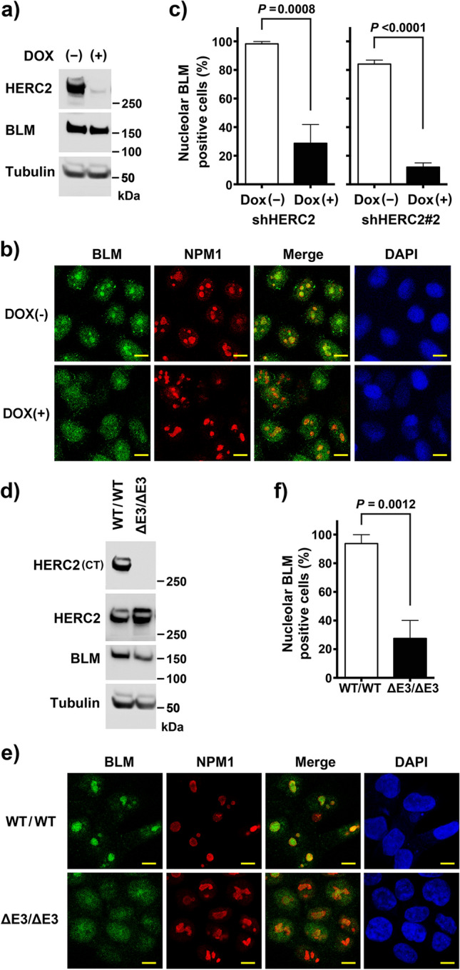
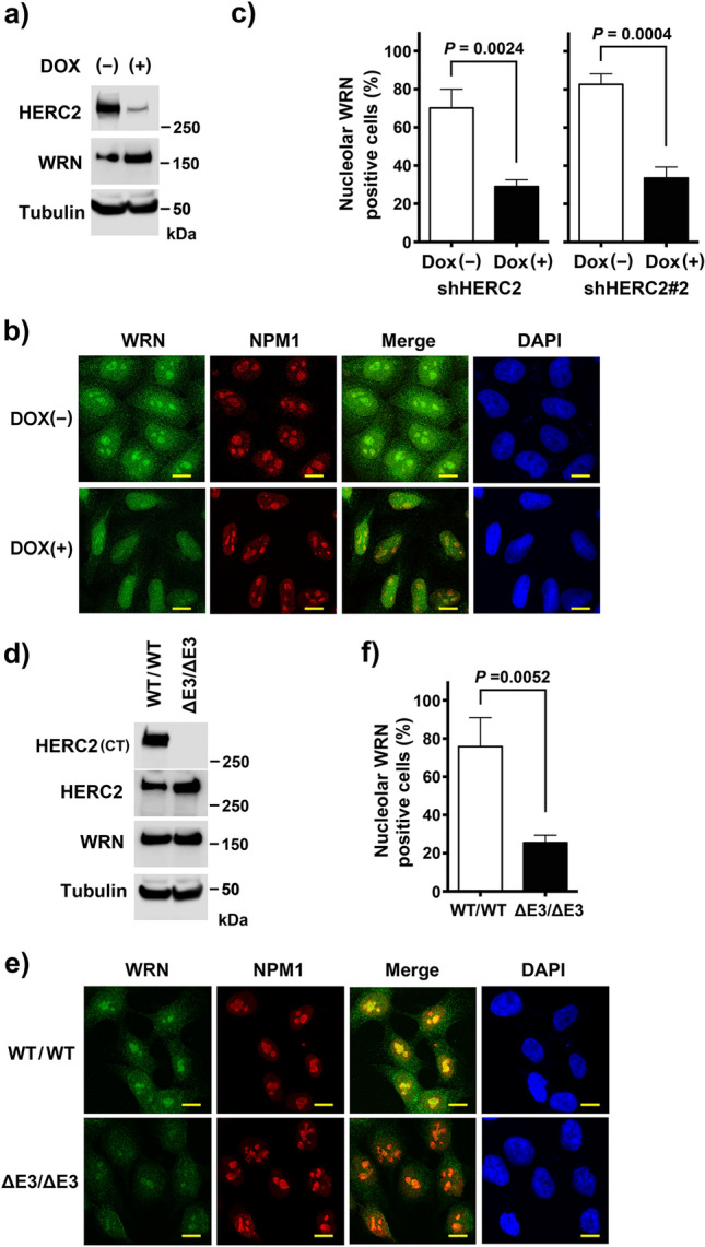
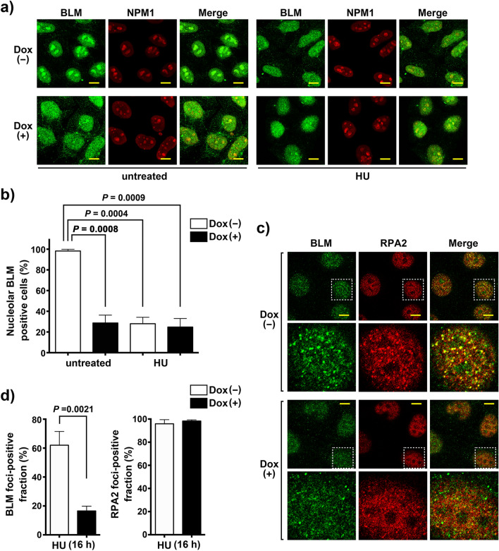
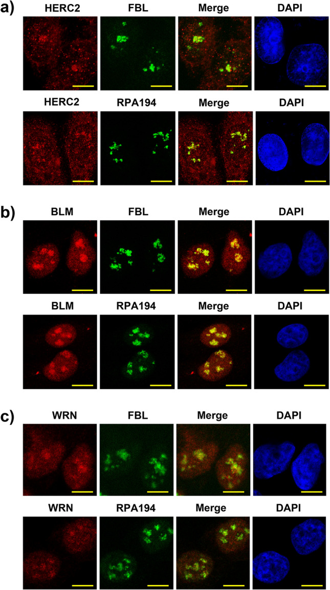
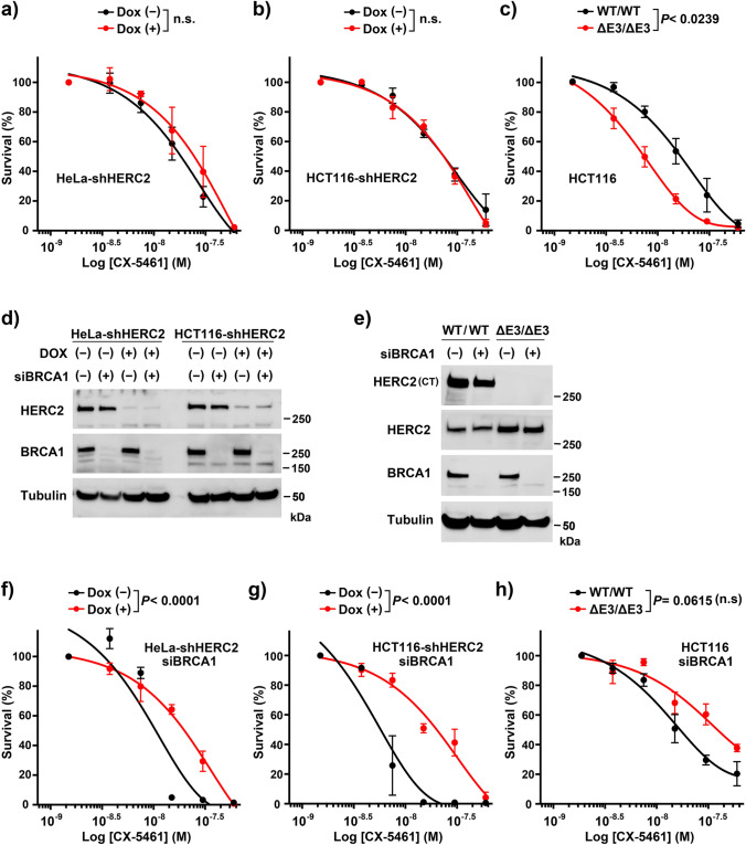
The nucleolus is a nuclear structure composed of ribosomal DNA (rDNA), and functions as a site for rRNA synthesis and processing. The rDNA is guanine-rich and prone to form G-quadruplex (G4), a secondary structure of DNA. We have recently found that HERC2, an HECT ubiquitin ligase, promotes BLM and WRN RecQ DNA helicases to resolve the G4 structure. Here, we report the role of HERC2 in the regulation of nucleolar localization of the helicases. Furthermore, HERC2 inactivation enhances the effects of CX-5461, an inhibitor of RNA polymerase I (Pol I)-mediated transcription of rRNA with an intrinsic G4-stabilizing activity. HERC2 depletion or homozygous deletion of the C-terminal HECT domain of HERC2 prevented the nucleolar localization of BLM and WRN, and inhibited relocalization of BLM to replication stress-induced nuclear RPA foci. HERC2 colocalized with fibrillarin and Pol I subunit RPA194, both of which are required for rRNA transcription. The HERC2 dysfunction enhanced the suppression of pre-rRNA transcription by CX-5461. These results suggest the effect of HERC2 status on the functions of BLM and WRN on rRNA transcription in the nucleolus. Since HERC2 is downregulated in numerous cancers, this effect may be clinically relevant considering the beneficial effects of CX-5461 in cancer treatments.


Conflict of interest statement

The authors declare no competing interests.

Figures

Figure 1
Figure 1
Nucleolar localization of BLM requires HERC2 and its E3 domain. (a,b) HeLa-shHERC2 cells, with or without Dox-mediated induction, were subjected to immunoblotting (a) or immunostain (b) with the indicated antibodies. The nuclei were counter stained with DAPI. Scale bar, 10 μm. (c) Quantifications of the nucleolar BLM positive cell from (b) (left panel), and those from HeLa cells with a different shRNA (shHERC2#2, right panel, see also Supplementary Figure S1) are shown. Error bars, S.D. of three independent experiments, each based on more than 100 cells. Statistical significances was calculated using the Student’s t-test. (d,e) Wild-type or HERC2ΔE3/ΔE3 HCT116 cells were subjected to immunoblotting (d) or immunostaining (e) with the indicated antibodies. Antibodies HERC2 (CT) and HERC2 recognize residues 4389–4834 and 1781–1974 of HERC2, respectively. Scale bar, 10 μm. (f) Quantification of the nucleolar BLM positive cell. Error bars, S.D. of three independent experiments, each based on more than 100 cells. Statistical significances was calculated using the Student’s t-test. Full-length blots/gels are presented in Supplementary Figure S12.
Figure 2
Figure 2
Nucleolar localization of WRN requires HERC2 and its E3 domain. (a,b) HeLa-shHERC2 cells, with or without Dox-mediated induction, were subjected to immunoblotting (a) or immunostain (b) with the indicated antibodies. The nuclei were counter stained with DAPI. Scale bar, 10 μm. (c) Quantifications of the nucleolar WRN positive cell from (b) (left panel), and those from HeLa cells with a different shRNA (shHERC2#2, right panel, see also Supplementary Figure S1) are shown. Error bars, S.D. of three independent experiments, each based on more than 100 cells. Statistical significances was calculated using the Student’s t-test. (d,e) Wild-type or HERC2ΔE3/ΔE3 HCT116 cells were subjected to immunoblotting (d) or immunostain (e) with the indicated antibodies. Scale bar, 10 μm. (f) Quantification of the nucleolar WRN positive cell. Error bars, S.D. of three independent experiments, each based on more than 100 cells. Statistical significances was calculated using the Student’s t-test. Full-length blots/gels are presented in Supplementary Figure S12.
Figure 3
Figure 3
BLM translocation in response to replication stress is dismissed by HERC2 depletion. (a) BLM disappears from the nucleolus in response to replication stress. HeLa-shHERC2 cells with or without Dox-mediated induction, untreated or treated with 1 mM HU for 4 h, were immunostained with antibodies against BLM and NPM1. Scale bar, 10 μm. (b) Quantification of the nucleolar BLM positive cell with or without HU treatment. Error bars, S.D. of three independent experiments, each based on more than 100 cells. Statistical significances was calculated using the Student’s t-test. (c) HERC2 is required for relocalization of BLM to RPA2 nuclear foci in response to replication stress. HeLa-shHERC2 cells with or without Dox-mediated induction, treated with 1 mM HU for 16 h, were immunostained with the indicated antibodies. One representative nucleus (dashed line) in the upper panels has been magnified and shown in the lower panels. Colocalization of BLM and RPA2 is shown in the merged display as yellow foci. Scale bar, 10 μm. (d) Quantification of the cells displaying more than ten BLM foci co-localizing with RPA2 (left panel) or discrete RPA2 foci (right panel). Error bars, S.D. of three independent experiments, each based on more than 50 cells. Statistical significances were calculated using Student’s t-test.
Figure 4
Figure 4
HERC2, BLM and WRN co-localize with FBL and RPA194. Spontaneously growing HeLa cells are subjected to immunostaining with HERC2 (a), BLM (b) or WRN (c) coimmunostained with FBL (upper panels) or RPA194 (lower panels) as indicated. The nuclei were counter stained with DAPI. Scale bar, 10 μm.
Figure 5
Figure 5
HERC2 dysfunction enhances the inhibitory effect of CX-5461 on pre-rRNA transcription. HeLa-shHERC2 (a) or HCT116-shHERC2 (b) cells untreated (solid line) or treated with Dox (dashed line), and wild-type (solid line) or HERC2ΔE3/ΔE3 (dashed line) HCT116 cells (c) were incubated with the indicated dose of CX5461. Inhibition of pre-rRNA (red) and c-Myc mRNA (black) transcription was analyzed by qRT-PCR, and was normalized against β-actin mRNA expression levels (see Supplementary Figure S8 for the expression level of β-actin mRNA). Data are shown as the means ± S.D of three independent experiments. P-values of interactions were calculated using two-way ANOVA. The concentration that inhibited 50% of the RNA products (IC50 value) was as follows: (a) pre-rRNA Dox (−): 0.77 μmol/l, Dox ( +): 0.21 μmol/l, others: n.a., (b) c-Myc-mRNA Dox (−): 0.47 μmol/l, Dox ( +): 0.52 μmol/l, pre-rRNA Dox (−): 0.26 μmol/l, Dox ( +) 0.06 μmol/l, (c) c-Myc-mRNA WT/WT: 0.15 μmol/l, ΔE3/ΔE3: 0.35 μmol/l, pre-rRNA WT/WT: 0.13 μmol/l, ΔE3/ΔE3 0.03 μmol/l.
Figure 6
Figure 6
HERC2 dysfunction alters the sensitivity of cells to CX-5461. (a–c) HeLa-shHERC2 (a) and HCT116-shHERC2 (b) cells, with or without Dox-mediated induction, or WT or HERC2ΔE3/ΔE3 HCT116 cells (c) were exposed to indicated doses of the CX5461 for 24 h and analyzed for clonogenic survival after two weeks. The data are shown with the nonlinear regression fit curves of one phase decay (GraphPad Prism). (d–h) Cells as in (a–c) were transfected with control (–) or BRCA1-specific siRNA, with or without Dox-mediated induction, and either subjected to immunoblotting with the indicated antibodies (d,e) or clonogenic survival assays (f–h, and supplementary Figure S11) as in (a–c). Average ± SD values, normalized to cells without agents were derived from three independent experiments. P-values of interactions were calculated using two-way ANOVA. The concentration that inhibited 50% of the colonies (IC50 value) was as follows: (a) Dox (−): 17.3 nmol/l, Dox ( +): 22.3 nmol/l, (b) Dox (−): 22.4 nmol/l, Dox ( +): 21.6 nmol/l, (c) WT/WT: 16.1 nmol/l, ΔE3/ΔE3: 7.3 nmol/l, (f) Dox (−): 10.0 nmol/l, Dox ( +): 18.3 nmol/l, (g) Dox (−): 6.1 nmol/l, Dox ( +): 18.1 nmol/l, (h) WT/WT: 17.8 nmol/l, ΔE3/ΔE3: 39.3 nmol/l. Full-length blots/gels are presented in Supplementary Figure S12.

Similar articles

Cited by

References

    1. Németh A, Längst G. Genome organization in and around the nucleolus. Trends. Genet. 2011;27:149–156. doi: 10.1016/j.tig.2011.01.002. - DOI - PubMed
    1. Boisvert FM, van Koningsbruggen S, Navascues J, Lamond AI. The multifunctional nucleolus. Nat. Rev. Mol. Cell. Biol. 2007;8:574–585. doi: 10.1038/nrm2184. - DOI - PubMed
    1. Lindström MS, et al. Nucleolus as an emerging hub in maintenance of genome stability and cancer pathogenesis. Oncogene. 2018;37:2351–2366. doi: 10.1038/s41388-017-0121-z. - DOI - PMC - PubMed
    1. Ogawa LM, Baserga SJ. Crosstalk between the nucleolus and the DNA damage response. Mol. Biosyst. 2017;13:443–455. doi: 10.1039/C6MB00740F. - DOI - PMC - PubMed
    1. Weeks SE, Metge BJ, Samant RS. The nucleolus: a central response hub for the stressors that drive cancer progression. Cell. Mol. Life. Sci. 2019;76:4511–4524. doi: 10.1007/s00018-019-03231-0. - DOI - PMC - PubMed

Publication types

LinkOut - more resources