

A novel quantitative and reference-free ultrasound analysis to discriminate different concentrations of bone mineral content
- PMID: 33432022
- PMCID: PMC7801603
- DOI: 10.1038/s41598-020-79365-0
A novel quantitative and reference-free ultrasound analysis to discriminate different concentrations of bone mineral content
Abstract
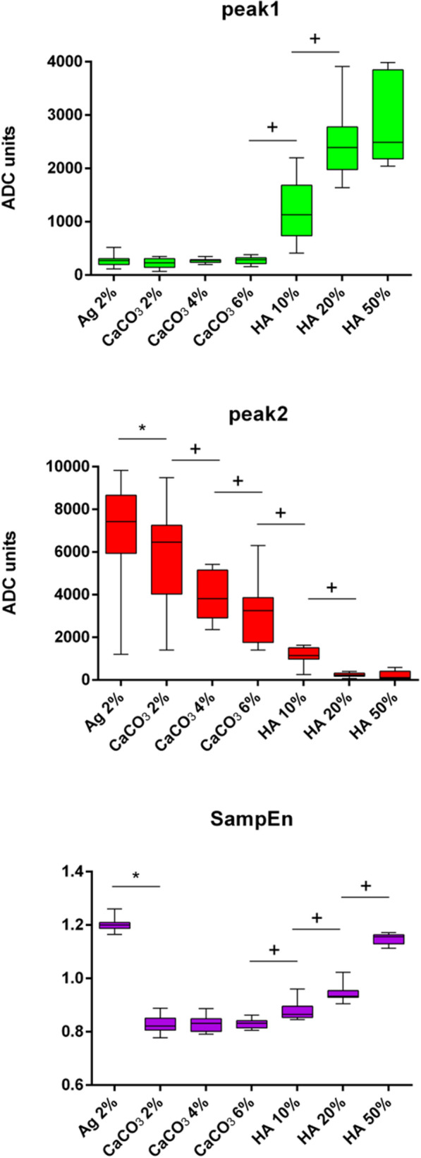
Bone fracture is a continuous process, during which bone mineral matrix evolves leading to an increase in hydroxyapatite and calcium carbonate content. Currently, no gold standard methods are available for a quantitative assessment of bone fracture healing. Moreover, the available tools do not provide information on bone composition. Whereby, there is a need for objective and non-invasive methods to monitor the evolution of bone mineral content. In general, ultrasound can guarantee a quantitative characterization of tissues. However, previous studies required measurements on reference samples. In this paper we propose a novel and reference-free parameter, based on the entropy of the phase signal calculated from the backscattered data in combination with amplitude information, to also consider absorption and scattering phenomena. The proposed metric was effective in discriminating different hydroxyapatite (from 10 to 50% w/v) and calcium carbonate (from 2 to 6% w/v) concentrations in bone-mimicking phantoms without the need for reference measurements, paving the way to their translational use for the diagnosis of tissue healing. To the best of our knowledge this is the first time that the phase entropy of the backscattered ultrasound signals is exploited for monitoring changes in the mineral content of bone-like materials.
Conflict of interest statement
The authors declare no competing interests.
Figures


Similar articles
-
Quantitative imaging of ultrasound backscattered signals with information entropy for bone microstructure characterization.Sci Rep. 2022 Jan 10;12(1):414. doi: 10.1038/s41598-021-04425-y. Sci Rep. 2022. PMID: 35013540 Free PMC article.
-
Liquid Calibration Phantoms in Ultra-Low-Dose QCT for the Assessment of Bone Mineral Density.J Clin Densitom. 2020 Jan-Mar;23(1):108-116. doi: 10.1016/j.jocd.2019.02.005. Epub 2019 Feb 22. J Clin Densitom. 2020. PMID: 30902572
-
Bone-composition imaging using coherent-scatter computed tomography: assessing bone health beyond bone mineral density.Med Phys. 2006 Apr;33(4):904-15. doi: 10.1118/1.2179151. Med Phys. 2006. PMID: 16696465
-
The Role of Matrix Composition in the Mechanical Behavior of Bone.Curr Osteoporos Rep. 2018 Jun;16(3):205-215. doi: 10.1007/s11914-018-0433-0. Curr Osteoporos Rep. 2018. PMID: 29611037 Free PMC article. Review.
-
Quantitative ultrasound: a technique to target women with low bone mass for preventive therapy.Am J Med. 1995 Feb 27;98(2A):48S-51S. doi: 10.1016/s0002-9343(05)80046-4. Am J Med. 1995. PMID: 7709935 Review.
Cited by
-
The progress in quantitative evaluation of callus during distraction osteogenesis.BMC Musculoskelet Disord. 2022 May 24;23(1):490. doi: 10.1186/s12891-022-05458-8. BMC Musculoskelet Disord. 2022. PMID: 35610718 Free PMC article. Review.
-
A Systematized Review of Quantitative Ultrasound Based on First-Order Speckle Statistics.IEEE Trans Ultrason Ferroelectr Freq Control. 2024 Jul;71(7):872-886. doi: 10.1109/TUFFC.2024.3402560. Epub 2024 Jul 9. IEEE Trans Ultrason Ferroelectr Freq Control. 2024. PMID: 38758626 Free PMC article.
-
Ultrasound Imaging in Knee Osteoarthritis: Current Role, Recent Advancements, and Future Perspectives.J Clin Med. 2024 Aug 21;13(16):4930. doi: 10.3390/jcm13164930. J Clin Med. 2024. PMID: 39201072 Free PMC article. Review.
-
Ultrasound identification of the cementoenamel junction and clinical correlation through ex vivo analysis.Sci Rep. 2024 Nov 13;14(1):27821. doi: 10.1038/s41598-024-79081-z. Sci Rep. 2024. PMID: 39537843 Free PMC article.
-
Quantitative ultrasound classification of healthy and chemically degraded ex-vivo cartilage.Sci Rep. 2025 Jul 1;15(1):20760. doi: 10.1038/s41598-025-07827-4. Sci Rep. 2025. PMID: 40596644 Free PMC article.
References
-
- Mamou J, Oelze ML. Quantitative ultrasound in soft tissues. Quant. Ultrasound Soft Tissues. 2013 doi: 10.1007/978-94-007-6952-6. - DOI
Publication types
MeSH terms
LinkOut - more resources
Full Text Sources
Other Literature Sources
Medical
