. 2021 Jan 11;11(1):413.
doi: 10.1038/s41598-020-80464-1.

No evidence for basigin/CD147 as a direct SARS-CoV-2 spike binding receptor

Affiliations

No evidence for basigin/CD147 as a direct SARS-CoV-2 spike binding receptor

Jarrod Shilts et al. Sci Rep. .

Abstract

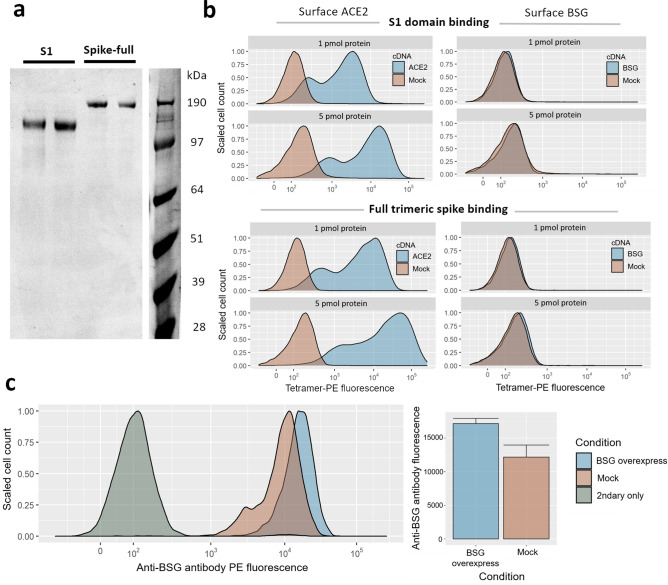
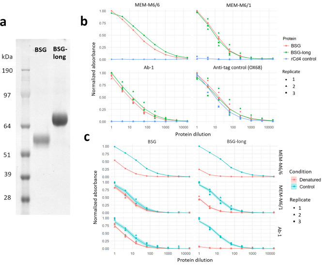
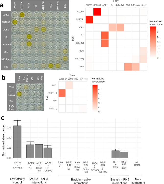
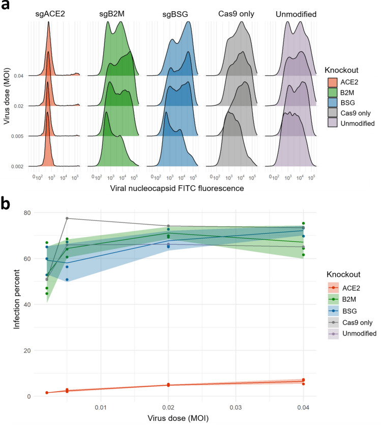
The spike protein of SARS-CoV-2 is known to enable viral invasion into human cells through direct binding to host receptors including ACE2. An alternate entry receptor for the virus was recently proposed to be basigin/CD147. These early studies have already prompted a clinical trial and multiple published hypotheses speculating on the role of this host receptor in viral infection and pathogenesis. Here, we report that we are unable to find evidence supporting the role of basigin as a putative spike binding receptor. Recombinant forms of the SARS-CoV-2 spike do not interact with basigin expressed on the surface of human cells, and by using specialized assays tailored to detect receptor interactions as weak or weaker than the proposed basigin-spike binding, we report no evidence for a direct interaction between the viral spike protein to either of the two common isoforms of basigin. Finally, removing basigin from the surface of human lung epithelial cells by CRISPR/Cas9 results in no change in their susceptibility to SARS-CoV-2 infection. Given the pressing need for clarity on which viral targets may lead to promising therapeutics, we present these findings to allow more informed decisions about the translational relevance of this putative mechanism in the race to understand and treat COVID-19.


Conflict of interest statement

The authors declare no competing interests.

Figures

Figure 1
Figure 1
Gain of SARS-CoV-2 spike binding activity on human cells over-expressing ACE2 but not BSG. (a) Expression and purification of the S1 domain and full ectodomain of the SARS-CoV-2 spike protein produced in human cell lines. Two independent preparations of purified spike were resolved by SDS-PAGE under reducing conditions and stained with Coomassie blue dye. (b) Cells transfected with cDNAs encoding ACE2 but not BSG bind highly avid fluorescent SARS-CoV-2 spike tetramers. Flow cytometry fluorescence distributions of cells stained with tetramers composed of biotinylated spike protein either using the S1 domain (top panels) or the entire ectodomain (lower panels) clustered around phycoerythrin-conjugated streptavidin. The stained HEK293 cells were transfected with cDNA to overexpress either ACE2 (left) or BSG (right). Mock-transfected cells are shown in red. Similar behavior to the data shown was observed in three separate tests. (c) Transfection with BSG cDNA leads to upregulation of cell-surface BSG. Surface basigin levels on HEK293 cells labeled with an anti-human BSG monoclonal antibody. BSG levels are compared to a negative control of secondary-antibody only. Mean antibody fluorescence intensities are summarized in the adjacent bar graph, with error bars showing standard deviations (n = 2–4).
Figure 2
Figure 2
Basigin expressed as recombinant protein ectodomains retain their biochemical activity. (a) Expression and purification of two and three Ig-like domain isoforms of basigin. Proteins were resolved under reducing conditions by SDS-PAGE and stained with a Coomassie dye (b). Recombinant basigin but not control proteins are recognized by anti-basigin monoclonal antibodies. ELISA dilution series of BSG and BSG-long recognized by three different monoclonal antibodies, and a control OX68 antibody against their tags. A negative control of a recombinant Cd4 tag is included for each antibody. (c) Recombinant basigin retains folded conformation of epitopes recognized by three different monoclonal antibodies. ELISA dilution curves comparing unmodified basigin to protein treated with heat and a reducing agent. Three replicates were performed for all ELISA curves except MEM-M6/6, for which only a single trial was done. Dose response curve model fit lines are superimposed on the data points, with shading indicating the 95% confidence bounds of the models.
Figure 3
Figure 3
Sensitive assays designed to detect extracellular protein interactions do not detect a direct interaction between human basigin and the SARS-CoV-2 spike protein. (a) No signs of spike-basigin binding in an avidity-based protein interaction assay systematically testing a matrix of recombinant baits immobilized to streptavidin-coated plates (rows) against preys clustered around HRP-conjugated streptavidin (columns). A photograph of a representative assay plate (left) is shown alongside background-corrected absorbance values averaged across two replicates. (b) The emerging D614G mutant variant of the SARS-CoV-2 spike also does not bind basigin. Binding matrix including the common D614G variant of the SARS-CoV-2 spike protein instead of the reference sequence. (c) Spike protein binding to basigin is consistently undetectable compared to other control interactions. Binding signals were averaged across bait and prey orientations for known interacting protein pairs, the basigin-spike pairs, and all other pairs. Error bars represent standard deviation from the mean (n = 2, except “All others” n = 58).
Figure 4
Figure 4
Knockdown of basigin by CRISPR-Cas9 in a lung cell line has no effect on susceptibility to SARS-CoV-2 infection. (a) Knockout of the known viral entry receptor ACE2 blocks viral infection in CaLu-3 cells, but not single guide (sgRNA) knockouts of basigin or negative control MHC class I receptors. Representative flow-cytometry profiles of SARS-CoV-2 nucleocapsid protein in CaLu-3 cells 48 h post-infection. (b) Lung cells with cell-surface basigin knocked down have no significant change in rates of viral entry and SARS-CoV-2 infection. Three replicates were performed of all 3 knockout conditions. Shading represents standard deviation from the mean percent of cells infected.

References

    1. Hoffmann M, et al. SARS-CoV-2 cell entry depends on ACE2 and TMPRSS2 and is blocked by a clinically proven protease inhibitor. Cell. 2020 doi: 10.1016/j.cell.2020.02.052. - DOI - PMC - PubMed
    1. Lan J, et al. Structure of the SARS-CoV-2 spike receptor-binding domain bound to the ACE2 receptor. Nature. 2020 doi: 10.1038/s41586-020-2180-5. - DOI - PubMed
    1. Wrapp D, et al. Cryo-EM structure of the 2019-nCoV spike in the prefusion conformation. Science. 2020;367:1260–1263. doi: 10.1126/science.abb2507. - DOI - PMC - PubMed
    1. Chan C-M, et al. Carcinoembryonic antigen-related cell adhesion molecule 5 is an important surface attachment factor that facilitates entry of middle east respiratory syndrome coronavirus. J. Virol. 2016;90:9114–9127. doi: 10.1128/JVI.01133-16. - DOI - PMC - PubMed
    1. Chu H, et al. Middle East respiratory syndrome coronavirus and bat coronavirus HKU9 both can utilize GRP78 for attachment onto host cells. J. Biol. Chem. 2018;293:11709–11726. doi: 10.1074/jbc.RA118.001897. - DOI - PMC - PubMed

Publication types