. 2021 May;39(5):567-577.
doi: 10.1038/s41587-020-00781-8. Epub 2021 Jan 11.

Targeting herpes simplex virus with CRISPR-Cas9 cures herpetic stromal keratitis in mice

Affiliations

Targeting herpes simplex virus with CRISPR-Cas9 cures herpetic stromal keratitis in mice

Di Yin et al. Nat Biotechnol. 2021 May.

Abstract

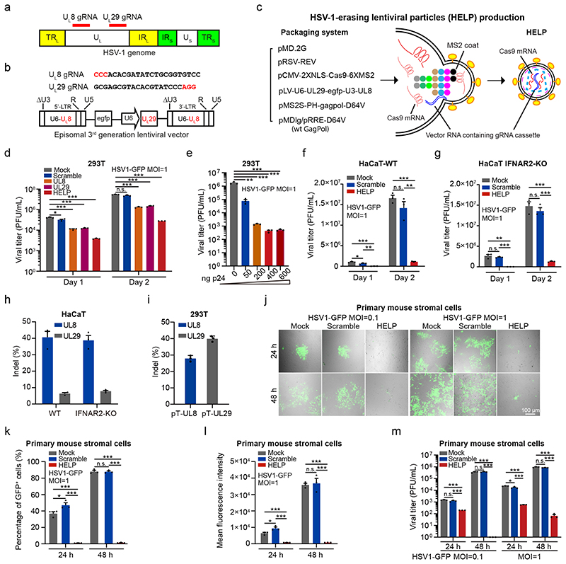
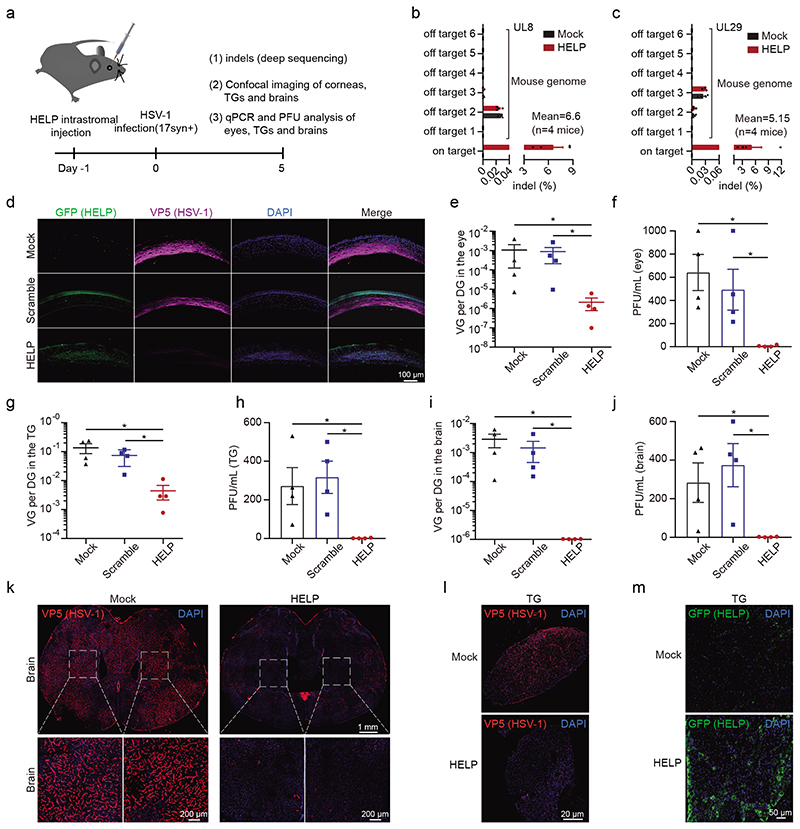
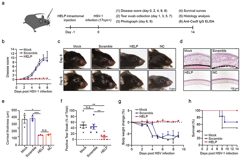
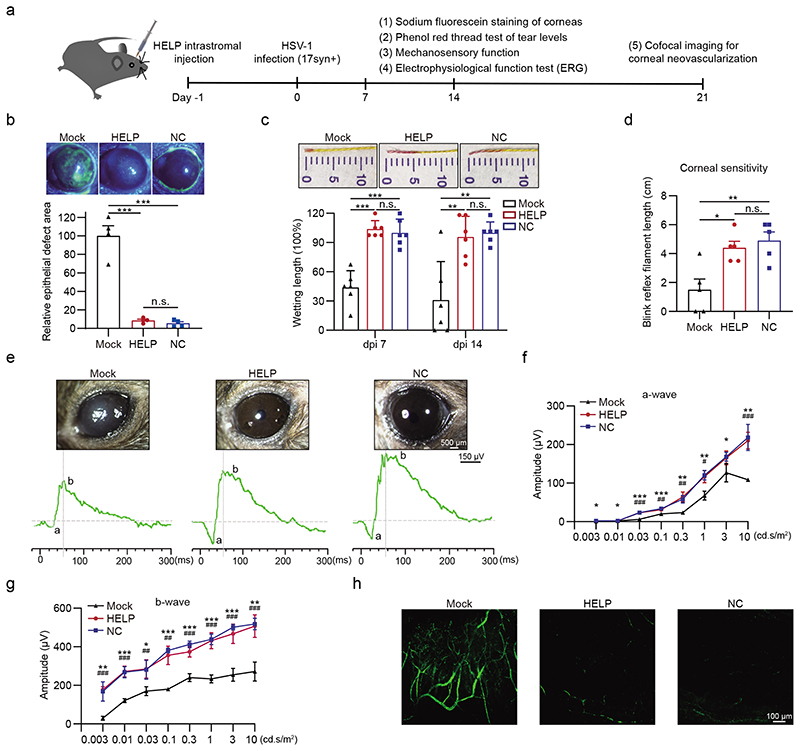
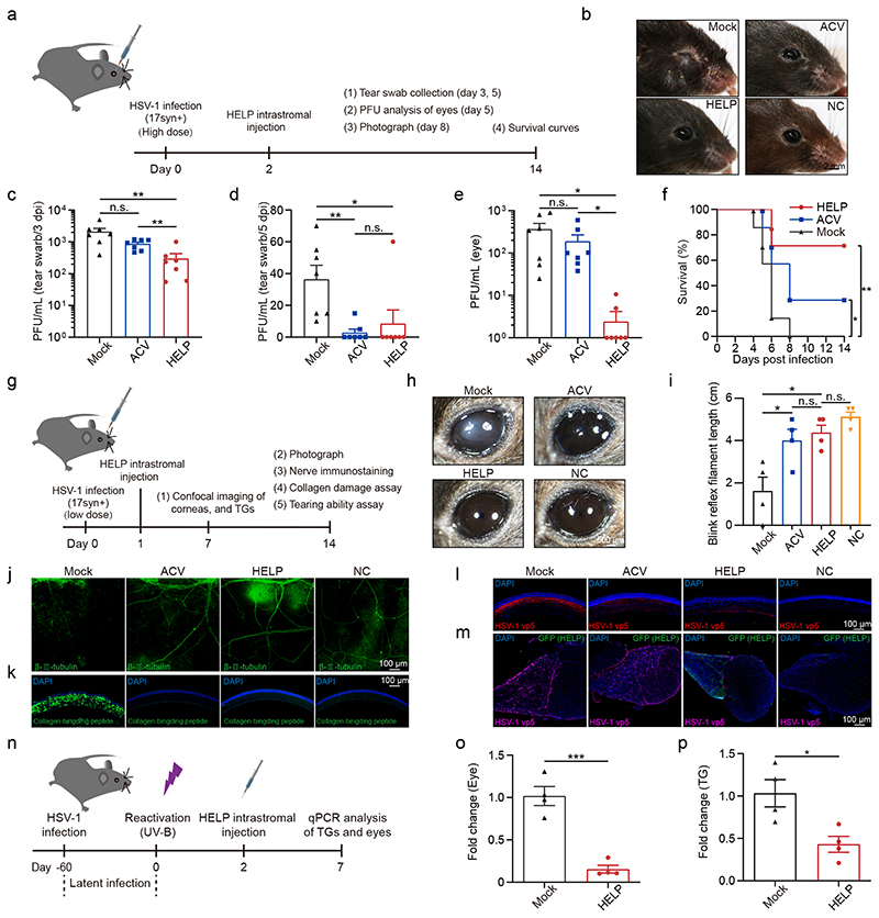
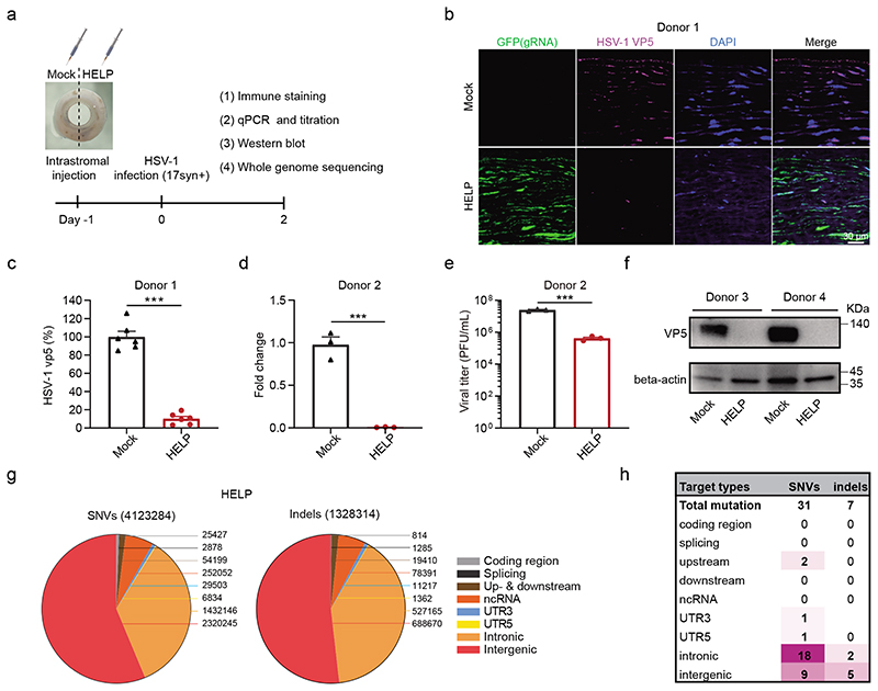
Herpes simplex virus type 1 (HSV-1) is a leading cause of infectious blindness. Current treatments for HSV-1 do not eliminate the virus from the site of infection or latent reservoirs in the trigeminal ganglia. Here, we target HSV-1 genomes directly using mRNA-carrying lentiviral particles that simultaneously deliver SpCas9 mRNA and viral-gene-targeting guide RNAs (designated HSV-1-erasing lentiviral particles, termed HELP). We show that HELP efficiently blocks HSV-1 replication and the occurrence of herpetic stromal keratitis (HSK) in three different infection models. HELP was capable of eliminating the viral reservoir via retrograde transport from corneas to trigeminal ganglia. Additionally, HELP inhibited viral replication in human-derived corneas without causing off-target effects, as determined by whole-genome sequencing. These results support the potential clinical utility of HELP for treating refractory HSK.


Conflict of interest statement

Competing interests

The authors declare no competing interests.

Figures

Figure 1
Figure 1. HELP blocks HSV-1 replication in vitro.
a, Schematic representation of the HSV-1 genome and gRNA loci. TRL, terminal repeat long; IRL, internal repeat long; UL, unique long; IRS, internal repeat short; TRS, terminal repeat short; US, unique short. b, The gRNA sequences and expression cassettes for HELP. c, Schematic illustration of HELP production. Coloured dots represent the main components of lentiviral Gag and GagPol polyproteins. Gag is composed of matrix (MA), capsid (CA), nucleocapsid (NC), whereas Pol consists of protease (PR), reverse transcriptase (RT) and integrase (IN). d-g, The anti-viral activity of HELP in cell lines. d, Mock vs. Scramble, UL8, UL29 and HELP, P=0.0220, 0.0003, 0.0003 and 0.0002, respectively, on day 1; P<0.0001 on day 2. e, Mock vs. 50 ng, P=0.0011; P=0.0009 for others. f, HELP vs. Mock and Scramble, P=0.0003 and 0.0019 on day 1; P<0.0001 and P=0.0011 on day 2. Mock vs. Scramble, P=0.0420 on day 1. g, HELP vs. Mock and Scramble, P=0.0013 and P<0.0001 on day 1; P=0.0002 and 0.0001 on day 2. h and i, TIDE analysis of indels in HSV1 genome and plasmid DNA. Viral DNA was from day 2 samples in f and g. j-m, The antiviral activity in primary mouse corneal stromal cells by confocal microscopy (j), flow cytometry (k and l) and PFU (m). k, Mock vs. HELP, P=0.0001 at 24 h; P<0.0001 for others. l, *P=0.0380, ***P=0.0005, ***P=0.0004, ***P<0.0001 and ***P=0.0003, left to right. m, ***P<0.0001, ***P=0.0010, ***P=0.0002, ***P<0.0001, *P=0.0236, ***P<0.0001, ***P=0.0006, ***P<0.0001, ***P<0.0001, left to right. Images are representative of three independent biological replicates in one experiment (j). Scale bars, 100 μm. The gating strategy is provided in Supplementary Fig. 20. Data and error bars represent mean ± SEM from three biologically independent replicates. Unpaired two-tailed Student’s t-tests. n.s.=non-significant.
Figure 2
Figure 2. HELP blocks HSV-1 infection of corneas and neurons in a prevention model.
a, Flow chart for evaluating antiviral effects of HELP in vivo. 100 ng p24 HELP, scramble mLP, or 2 μL PBS (Mock) were injected into corneas of mice by intrastromal injection. After 24 h, the mice were infected with HSV-1 17syn+ (2×10⁶ PFU/eye). b and c, Deep sequencing analysis of on-target effects in HSV-1 and off-target effects in the mouse genome. n=4 mice. d, Confocal imaging of HSV-1 and HELP in corneas. Mouse corneal sections were incubated with both anti-GFP (HELP) and anti-HSV-1 (VP5) antibodies. Scale bars, 100 μm. e-j, qPCR and PFU analysis of HSV-1 dissemination in the eyes, trigeminal ganglia and brains. The abundance of HSV-1 shown as the viral genome (VG) per diploid genome (DG). n=4 mice. P=0.0286 in e-j. k and l, Confocal analysis of HSV-1 presence in the whole brain and TG. Scale bars, 1 mm, 200 μm and 20 μm, respectively. m, Confocal analysis of HELP presence in the TG after intracorneal injection. Scale bars, 50 μm. Data and error bars represent mean ± SEM. Unpaired two-tailed Mann-Whitney-tests. The experiments were repeated twice with similar results.
Figure 3
Figure 3. HELP suppresses HSV-1-associated disease pathologies in the prevention model.
a, Flow chart for evaluating antiviral effects of HELP in vivo. 100 ng p24 HELP, scramble mLP or 2 μL PBS (Mock) were injected into corneas. After 24 h, the mice were infected with HSV-1 17syn+ (2×10⁶ PFU/eye). b, Ocular disease scores (0 to 4, 4 being severe) in mice. n=6 mice. c, Photographs of the right eyes of differently treated mice on 6 dpi and 9 dpi. Each image is a representative of 3 mice in one experiment. NC, non-treated control. Scale bars, 2 mm. d, Corneal histology of eyes on 14 dpi. Each image is a representative of 3 mice in two independent experiments (c, d). Scale bars, 100 μm. e, Thickness of the corneas assessed from histology. n=3 mice. HELP vs. Mock and Scramble, P=0.0168 and 0.0006. f, Secreted HSV-1 assessed from the swabs of eyes. Tear swabs from each mouse was collected at 1, 3, 5, 7 dpi during the experiment. The percentage of HSV-1 positive swabs was recorded. n=6 mice. Mock vs. HELP, P=0.0056; Scramble vs. HELP, P=0.0072. g, Bodyweight. n=6 mice. h, Kaplan-Meier survival curves. n=6 mice. Data and error bars represent mean ± SEM. Unpaired two-tailed Student’s t-tests. n.s.=non-significant.
Figure 4
Figure 4. Eye health after HELP treatment in the prevention model.
a, Flow chart for evaluating eye health in the prevention model. 100 ng p24 HELP or 2 μL PBS (Mock) were delivered into corneas 24 h before the mice were infected with HSV-1 17syn+ (2×10⁶ PFU/eye). b, Sodium fluorescein staining of mice corneas. The defect area of HELP and NC was normalized to Mock. n=4 mice. Mock vs. HELP and NC, P=0.0002 and 0.0001. c, Phenol red thread test of the wettability of the tear fluid. n=6 mice. Mock vs. HELP, P<0.0001 on dpi 7 and P=0.0054 on dpi 14; Mock vs. NC, P=0.0001 on dpi 7 and P=0.0021 on dpi 14. d, Measuring the mechanosensory function of the corneas by esthesiometer. n=5 mice. Mock vs. HELP, P=0.0104; Mock vs. NC, P=0.0074. e-g, Change in ERG amplitudes of treated eyes. Corneal graphs and traces of the a-waves and b-waves (e). Quantitative analysis of a-wave and b-wave amplitude (f and g). n=5 mice. NC vs. Mock (*), P=0.02, 0.01, 0.00002, 0.0006, 0.0026, 0.0036, 0.03 and 0.002; HELP vs. Mock (#), P=0.0002, 0.0092, 0.0018, 0.0114 and 0.0005 for ascending cd.s/m2(f). NC vs. Mock (*), P=0.0040, 0.0003, 0.0200, 0.00001, 0.00001, 0.0001, 0.0001 and 0.0015; HELP vs. Mock (#), P=0.0001, 0.0002, 0.0047, 0.0011, 0.0007, 0.0004, 0.0005 and 0.0005 for ascending cd.s/m2(g). h, Confocal microscopy imaging of neovascularization in the corneas. Data and error bars represent mean ± SEM. Unpaired two-tailed Student’s t-tests. n.s.=non-significant. Each image is a representative of 3 mice in one experiment (b, e, h).
Figure 5
Figure 5. HELP cures HSK in the therapeutic and recurrent models.
a, Flow chart for evaluating the antiviral effects of HELP in the HSK therapeutic model. Mice were infected with HSV-1 17syn+ (2×10⁶ PFU/eye). After 48 h, 100 ng p24 HELP or 2 μL PBS (Mock) was administrated. ACV was added topically to both eyes every day for five days. b, Photographs of the eyes of differently treated mice. c and d, Infectious units in tear swabs on 3 dpi and 5 dpi. n=7 mice. c, Mock vs. HELP, P=0.0083; ACV vs. HELP, P=0.0060. d, Mock vs. ACV, P=0.0031; Mock vs. HELP, P=0.0431. e, Plaque assay for HSV-1 in the eyes. n=7 mice. Mock vs. HELP, P=0.0197; ACV vs. HELP, P=0.0360. f, Kaplan-Meier survival curves. n=6 mice. Mock vs. HELP and ACV, P=0.0076 and 0.0297. g, Flow chart for evaluating the antiviral effects of HELP in the HSK therapeutic model. HSV-1 was used 5×10⁴ PFU/eye. h, Photographs of the differently treated eyes. Scale bars, 500 μm. i, Measuring the mechanosensory function of the corneas by esthesiometer. n=4 mice. Mock vs. HELP and ACV, P=0.0127 and 0.0349. j and k, Confocal images of sensory fibers and damaged collagen in corneas. Scale bars, 100 μm. l and m, Confocal images of HSV-1 (VP5) in the corneas and HELP (anti-GFP) in the TG. Scale bars, 100 μm. n, Flow chart for evaluating antiviral activity using a recurrent HSK model. Mice were infected with HSV-1 17syn+ (2×10⁵ PFU/eye). o and p, Virus load in the eyes and TGs detected by qPCR. n=4 mice. o, P=0.0004; p, P=0.0179. Data and error bars represent mean ± SEM. Unpaired two-tailed Student’s t-tests. n.s.=non-significant. Each image is a representative of 4 mice (b, h) or 2 mice (j-m) in one experiment.
Figure 6
Figure 6. HELP eliminates HSV-1 in tissue culture of human corneas.
a, Flow chart for evaluating antiviral effects of HELP in human corneas. 1.5 μg p24 HELP or 15 μL PBS (Mock) were injected into the corresponding punches derived from the same human cornea. After 24 h, the corneal punches were infected with HSV-1 17syn+ (2×10⁶ PFU/piece). b, Confocal analysis of the distribution of HSV-1 and HELP in the human cornea. GFP is indicative of the presence of HELP and VP5 for HSV-1. Scale bar, 30 μm. c, Percentage of VP5+ cells presented in (b). Data shown as the percentage of mock-treated tissues. n=6 biologically independent samples. P<0.0001. d, qPCR analysis the fold change of HSV-1 genome. n=3 biologically independent samples. P=0.0005. e, Titration of supernatants from human corneal cultures. n=3 biologically independent samples. P=0.0003. f, Western blot analysis of VP5 protein. The experiment was repeated twice with similar results. g, Identification of SNV and indel mutations in the HELP treated corneal punch at WGS level. Valid sequencing data were aligned to Human Genome version 19 (HG19). h, Summary of unique SNV and indel mutations. Data and error bars represent mean ± SEM. Unpaired two-tailed Student’s t-tests.

References

    1. Liesegang TJ. Herpes simplex virus epidemiology and ocular importance. Cornea. 2001;20:1–13. - PubMed
    1. Paludan SR, Bowie AG, Horan KA, Fitzgerald KA. Recognition of herpesviruses by the innate immune system. Nat Rev Immunol. 2011;11:143–154. - PMC - PubMed
    1. Bradshaw MJ, Venkatesan A. Herpes Simplex Virus-1 Encephalitis in Adults: Pathophysiology, Diagnosis, and Management. Neurotherapeutics. 2016;13:493–508. - PMC - PubMed
    1. Farooq AV, Shukla D. Herpes simplex epithelial and stromal keratitis: an epidemiologic update. Surv Ophthalmol. 2012;57:448–462. - PMC - PubMed
    1. Crumpacker CS, Schaffer PA. New anti-HSV therapeutics target the helicase–primase complex. Nat Med. 2002;8:327–328. - PubMed

Publication types

LinkOut - more resources