The use of chest ultrasonography in suspected cases of COVID-19 in the emergency department
- PMID: 33432268
- PMCID: PMC7745656
- DOI: 10.2144/fsoa-2020-0127
The use of chest ultrasonography in suspected cases of COVID-19 in the emergency department
Abstract
Aim: Severe acute respiratory syndrome coronavirus 2 (SARS-CoV-2) virus-specific reverse transcriptase-polymerase chain reaction (RT-PCR) represents the diagnostic gold standard. We explored the value of chest ultrasonography to predict positivity to SARS-CoV-2 on RT-PCR in suspected COVID-19 cases.
Patients & methods: Consecutive patients with suspect COVID-19 were included if they had fever and/or history of cough and/or dyspnea. Lung ultrasound score (LUSS) was computed according to published methods.
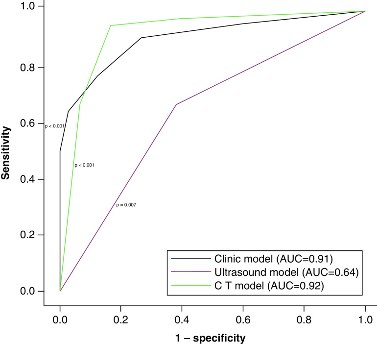
Results: A total of 76 patients were included. A 3-variable model based on aspartate transaminase (AST) > upper limit of normal, LUSS >12 and body temperature >37.5°C yielded an overall accuracy of 91%.
Conclusion: A simple LUSS-based model may represent a powerful tool for initial assessment in suspected cases of COVID-19.
Keywords: COVID-19; RT-PCR; SARS-CoV-2; lung ultrasound.
© 2020 Enrico Allegorico.
Conflict of interest statement
Financial & competing interests disclosure C Buonerba is a member of the Future Science OA Editorial Board. The authors have no other relevant affiliations or financial involvement with any organization or entity with a financial interest in or financial conflict with the subject matter or materials discussed in the manuscript apart from those disclosed. No writing assistance was utilized in the production of this manuscript.
Figures
Similar articles
-
Thoracic imaging tests for the diagnosis of COVID-19.Cochrane Database Syst Rev. 2020 Sep 30;9:CD013639. doi: 10.1002/14651858.CD013639.pub2. Cochrane Database Syst Rev. 2020. Update in: Cochrane Database Syst Rev. 2020 Nov 26;11:CD013639. doi: 10.1002/14651858.CD013639.pub3. PMID: 32997361 Updated.
-
Correlation between Chest Computed Tomography and Lung Ultrasonography in Patients with Coronavirus Disease 2019 (COVID-19).Ultrasound Med Biol. 2020 Nov;46(11):2918-2926. doi: 10.1016/j.ultrasmedbio.2020.07.003. Epub 2020 Jul 13. Ultrasound Med Biol. 2020. PMID: 32771222 Free PMC article.
-
Thoracic imaging tests for the diagnosis of COVID-19.Cochrane Database Syst Rev. 2020 Nov 26;11:CD013639. doi: 10.1002/14651858.CD013639.pub3. Cochrane Database Syst Rev. 2020. Update in: Cochrane Database Syst Rev. 2021 Mar 16;3:CD013639. doi: 10.1002/14651858.CD013639.pub4. PMID: 33242342 Updated.
-
Chest CT in the Emergency Department for Diagnosis of COVID-19 Pneumonia: Dutch Experience.Radiology. 2021 Feb;298(2):E98-E106. doi: 10.1148/radiol.2020203465. Epub 2020 Nov 17. Radiology. 2021. PMID: 33201791 Free PMC article.
-
Lung ultrasound score to monitor COVID-19 pneumonia progression in patients with ARDS.PLoS One. 2020 Jul 21;15(7):e0236312. doi: 10.1371/journal.pone.0236312. eCollection 2020. PLoS One. 2020. PMID: 32692769 Free PMC article.
Cited by
-
Ultrasound findings of lung ultrasonography in COVID-19: A systematic review.Eur J Radiol. 2022 Mar;148:110156. doi: 10.1016/j.ejrad.2022.110156. Epub 2022 Jan 20. Eur J Radiol. 2022. PMID: 35078136 Free PMC article.
-
Signs and symptoms to determine if a patient presenting in primary care or hospital outpatient settings has COVID-19.Cochrane Database Syst Rev. 2022 May 20;5(5):CD013665. doi: 10.1002/14651858.CD013665.pub3. Cochrane Database Syst Rev. 2022. PMID: 35593186 Free PMC article.
-
Utility and diagnostic test properties of pulmonary and cardiovascular point of care ultra-sonography (POCUS) in COVID-19 patients admitted to critical care unit.Eur J Radiol Open. 2022;9:100451. doi: 10.1016/j.ejro.2022.100451. Epub 2022 Nov 3. Eur J Radiol Open. 2022. PMID: 36345320 Free PMC article.
References
-
- Cevik M, Bamford C, Ho A. COVID-19 pandemic – a focused review for clinicians. Clin. Microbiol. Infect. S1198-743X(20)30231-7 (2020). https://pubmed.ncbi.nlm.nih.gov/32344166 - PMC - PubMed
LinkOut - more resources
Full Text Sources
Other Literature Sources
Miscellaneous