. 2021 Jul;79(1):67-76.
doi: 10.1111/his.14334. Epub 2021 Apr 14.

Molecular markers of telomere dysfunction and senescence are common findings in the usual interstitial pneumonia pattern of lung fibrosis

Affiliations

Molecular markers of telomere dysfunction and senescence are common findings in the usual interstitial pneumonia pattern of lung fibrosis

Joyce S Lee et al. Histopathology. 2021 Jul.

Abstract

Aims: Idiopathic pulmonary fibrosis (IPF) is a genetically mediated, age-associated, progressive form of pulmonary fibrosis characterised pathologically by a usual interstitial pneumonia (UIP) pattern of fibrosis. The UIP pattern is also found in pulmonary fibrosis attributable to clinical diagnoses other than IPF (non-IPF UIP), whose clinical course is similarly poor, suggesting common molecular drivers. This study investigates whether IPF and non-IPF UIP lungs similarly express markers of telomere dysfunction and senescence.

Methods and results: To test whether patients with IPF and non-IPF UIP share molecular drivers, lung tissues from 169 IPF patients and 57 non-IPF UIP patients were histopathologically and molecularly compared. Histopathological changes in both IPF and non-IPF UIP patients included temporal heterogeneity, microscopic honeycombing, fibroblast foci, and dense collagen fibrosis. Non-IPF UIP lungs were more likely to have lymphocytic infiltration, non-caseating granulomas, airway-centred inflammation, or small airways disease. Telomeres were shorter in alveolar type II (AECII) cells of both IPF and non-IPF UIP lungs than in those of age-similar, unused donor, controls. Levels of molecular markers of senescence (p16 and p21) were elevated in lysates of IPF and non-IPF UIP lungs. Immunostaining localised expression of these proteins to AECII cells. The mucin 5B (MUC5B) gene promoter variant minor allele frequency was similar between IPF and non-IPF UIP patients, and MUC5B expression was similar in IPF and non-IPF UIP lungs.

Conclusions: Molecular markers of telomere dysfunction and senescence are pathologically expressed in both IPF and non-IPF UIP lungs. These findings suggest that common molecular drivers may contribute to the pathogenesis of UIP-associated pulmonary fibrosis, regardless of the clinical diagnosis.

Keywords: alveolar type II cell; hypersensitivity pneumonitis; interstitial lung disease; pulmonary fibrosis; rheumatoid arthritis; scleroderma; senescence; telomere.


Figures

Figure. 1.
Figure. 1.. AECII cell telomere length quantification by teloFISH.
(A) Example of Cy3-labeled telomere signal (red), SPC–immunoreactive cells (green), and nuclei stained with DAPI (blue) in age similar unused donor control and (B) non-IPF UIP patient samples. (C) Telomere length was quantified by teloFISH on sections of lung harvested from age similar unused donor control, IPF and non-IPF UIP patient lung sections. Each data point represents average telomere fluorescence intensity in surfactant protein C (SPC)-immunoreactive cells for an individual patient. Magnification: ×40. Telomere fluorescence intensity was quantified with respect to DAPI area using Metamorph software. Data were collected from 11 IPF, 11non-IPF UIP (5 scleroderma, 1 rheumatoid arthritis, 4 hypersensitivity pneumonitis, 1 Sjogren’s syndrome) and 12 unused donor controls, **P < 0.01
Figure. 2.
Figure. 2.. P16 levels in IPF lung lysates.
(A) Immunoblots for p16 shows significantly higher levels of p16 in lung lysates of IPF patients (n=12) and non-IPF UIP patients (n=15; 8 scleroderma, 1 rheumatoid arthritis, 4 hypersensitivity pneumonitis, 2 Sjogren’s syndrome ) relative to those from unused donor control subjects (n=12). (B) Relative densitometric ratios of p16 to β-actin are presented by bar graphs (* p < 0.01, **p < 0.05, *** p < 0.01).
Figure. 3.
Figure. 3.. p21 levels in IPF lung lysates.
(A) Immunoblots for p21 shows significantly higher levels of p21 in lung lysates of IPF patients (n=19) and non-IPF UIP patients (n=14; 7 scleroderma, 1 rheumatoid arthritis, 4 hypersensitivity pneumonitis, 2 Sjogren’s syndrome) relative to those from unused donor control subjects (n=12). (B) Relative densitometric ratios of p21 to β-actin are presented by bar graphs (* p < 0.01, **p < 0.05 compared to normal controls).
Figure. 4.
Figure. 4.. Immunohistochemistry of IPF, non-IPF UIP, and normal lung with p16 or p21 antibodies.
Normal, IPF, and non-IPF UIP lungs were immunostained for p16 or co-immunostained for p21 (green fluorescence) and surfactant protein C (red fluorescence). IPF lungs stained with secondary antibodies alone (nonimmune) served as controls. Images are representative of tissues stained from 12 patients with IPF, 8 patients with non-IPF UIP (4 scleroderma, 2 rheumatoid arthritis, and 2 hypersensitivity pneumonitis. Note: representative non-IPF UIP images are from a patient with scleroderma UIP), and 10 control subjects.
Figure. 5.
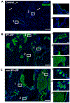
Figure. 5.. Immunohistochemistry of IPF, non-IPF UIP, and normal lung with MUC5B antibodies.
Lung tissue sections taken from 13 donor control (A), 14 IPF UIP (B) and 14 non IPF UIP (3 scleroderma, 4 rheumatoid arthritis, and 4 hypersensitivity pneumonitis, 1 Sjogren’s disease, 2 unclassifiable. Note: representative non-IPF UIP images are from a patient with scleroderma UIP). (C) were stained and examined for MUC5B (green) protein expression and distribution. Low power magnification image shows that in control lungs the majority of MUC5B was found in proximal (region 1, high power magnification on the right) airways, with some expression present in distal (region 2, high power magnification on the right) airways and alveoli (white arrows). Overall MUC5B expression was increased in IPF and non IPF UIP lungs and had similar distribution. Number of cells and amount of MUC5B per cell was increased in proximal (regions 1, high power magnification is on the right) and distal (regions 2, high power magnification is on the right) airways, with mucus accumulation in the lumen of the airways. Honeycomb cysts, lined with MUC5B expressing cells and with mucus accumulation in the lumen of the cysts, were present in both IPF and non IPF UIPs lungs. Nuclei were stained with DAPI (blue). Bar=500um for low power magnification, and 50um for high power magnification.

References

    1. Raghu G, Weycker D, Edelsberg J, Bradford WZ, Oster G. Incidence and prevalence of idiopathic pulmonary fibrosis. Am J Respir Crit Care Med. 2006;174(7):810–6. - PubMed
    1. Wolters PJ, Collard HR, Jones KD. Pathogenesis of idiopathic pulmonary fibrosis. Annual review of pathology. 2014;9:157–79. - PMC - PubMed
    1. Selman M, Lopez-Otin C, Pardo A. Age-driven developmental drift in the pathogenesis of idiopathic pulmonary fibrosis. Eur Respir J. 2016;48(2):538–52. - PubMed
    1. Alder JK, Chen JJ, Lancaster L, Danoff S, Su SC, Cogan JD, et al. Short telomeres are a risk factor for idiopathic pulmonary fibrosis. Proc Natl Acad Sci U S A. 2008;105(35):13051–6. - PMC - PubMed
    1. Kropski JA, Pritchett JM, Zoz DF, Crossno PF, Markin C, Garnett ET, et al. Extensive phenotyping of individuals at risk for familial interstitial pneumonia reveals clues to the pathogenesis of interstitial lung disease. Am J Respir Crit Care Med. 2015;191(4):417–26. - PMC - PubMed