. 2021 Aug;16(8):1582-1591.
doi: 10.4103/1673-5374.303036.

P2X7 receptor activation aggravates NADPH oxidase 2-induced oxidative stress after intracerebral hemorrhage

Affiliations

P2X7 receptor activation aggravates NADPH oxidase 2-induced oxidative stress after intracerebral hemorrhage

Hong Deng et al. Neural Regen Res. 2021 Aug.

Abstract

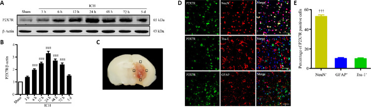
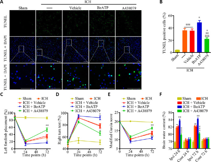
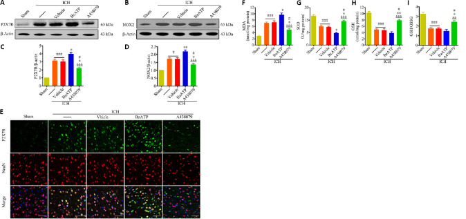
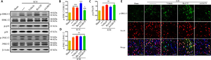
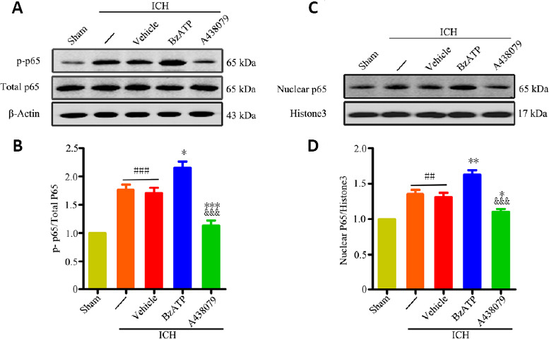
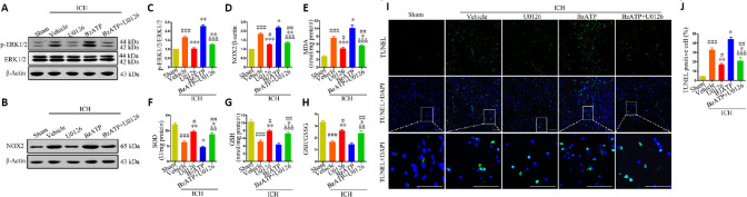
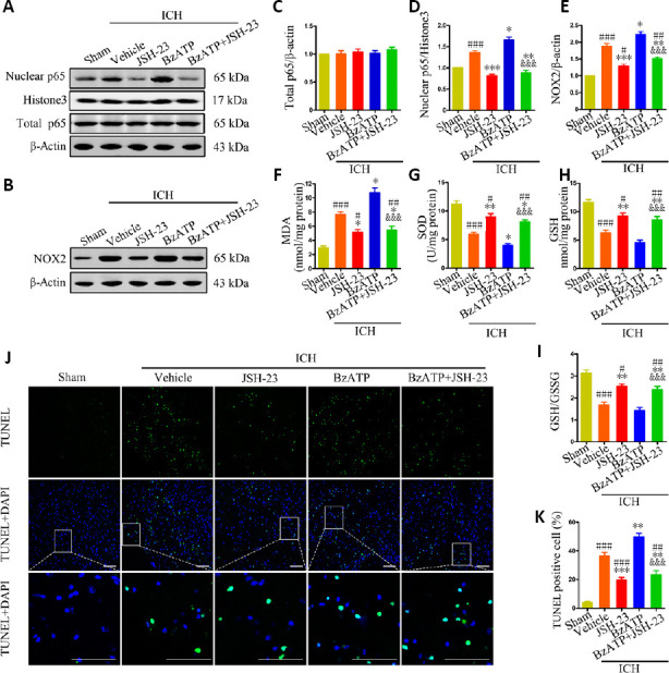
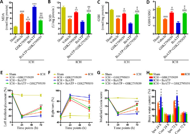
Oxidative stress is a crucial pathological process that contributes to secondary injury following intracerebral hemorrhage. P2X7 receptor (P2X7R), which is activated by the abnormal accumulation of extracellular ATP, plays an important role in the regulation of oxidative stress in the central nervous system, although the effects of activated P2X7R-associated oxidative stress after intracerebral hemorrhage remain unclear. Mouse models of intracerebral hemorrhage were established through the stereotactic injection of 0.075 U VII collagenase into the right basal ganglia. The results revealed that P2X7R expression peaked 24 hours after intracerebral hemorrhage, and P2X7R expressed primarily in neurons. The inhibition of P2X7R, using A438079 (100 mg/kg, intraperitoneal), reduced nicotinamide adenine dinucleotide phosphate oxidase 2 (NOX2) expression and malondialdehyde generation, increased superoxide dismutase and glutathione/oxidized glutathione levels, and alleviated neurological damage, brain edema, and apoptosis after intracellular hemorrhage. The P2X7R inhibitor A438079 (100 mg/kg, intraperitoneal injection) inhibited the activation of extracellular signal-regulated kinase 1/2 (ERK1/2) and nuclear factor kappa-B (NF-κB) after intracerebral hemorrhage. Blocking ERK1/2 activation, using the ERK1/2 inhibitor U0126 (2 µg, intraventricular injection), reduced the level of NOX2-mediated oxidative stress induced by P2X7R activation after intracellular hemorrhage. Similarly, the inhibition of NF-κB, using the NF-κB inhibitor JSH-23 (3.5 µg, intraventricular), reduced the level of NOX2-mediated oxidative stress induced by P2X7R activation. Finally, GSK2795039 (100 mg/kg, intraperitoneal), a NOX2 antagonist, attenuated P2X7R-mediated oxidative stress, neurological damage, and brain edema after intracerebral hemorrhage. The results indicated that P2X7R activation aggravated NOX2-induced oxidative stress through the activation of the ERK1/2 and NF-κB pathways following intracerebral hemorrhage in mice. The present study was approved by the Ethics Committee of Huazhong University of Science and Technology, China (approval No. TJ-A20160805) on August 26, 2016.

Keywords: brain; central nervous system; factor; inflammation; injury; pathways; repair; stroke.


Conflict of interest statement

None

Figures

Figure 1
Figure 1
Spatiotemporal expression of P2X7R in brain tissues near the hematoma after ICH. (A and B) Quantitative determination of P2X7R expression levels by western blot assay. The relative expression of P2X7R is expressed as the optical density ratio of P2X7R relative to that of β-actin. (C) Mouse brain slice showing the areas of brain tissues near the hematoma (indicated by black squares). These areas were examined by immunofluorescence and sampled for western blotting analysis. (D) P2X7R localization (green, stained by Alexa Fluor 488) was determined by immunofluorescence staining, 24 hours after ICH. The neuronal expression level of P2X7R was significantly higher than those observed for astrocytes and microglia. Iba-1, NeuN, and GFAP immunopositivity was detected using Cy3 (red). Scale bars: 50 µm. (E) Quantitative analysis of P2X7R expression in microglia (Iba-1+), neurons (NeuN+), and astrocytes (GFAP+). Data are presented as the mean ± SEM (n = 6). ###P < 0.001, vs. sham group; †††P < 0.001, vs. Iba-1+ or GFAP+ (one-way analysis of variance followed by Tukey’s post hoc test). GFAP: Glial fibrillary acidic protein; Iba-1: ionized calcium-binding adaptor molecule 1; ICH: intracerebral hemorrhage; P2X7R: P2X7 receptor.
Figure 2
Figure 2
P2X7R activation aggravates apoptosis, brain edema, and neurological impairments after ICH. (A) Apoptotic cells were detected by TUNEL staining, 24 hours after ICH. Low magnification, full-field and partial, high-magnification visual fields (indicated by the white frame) are displayed. The A438079 treatment group displayed a significantly reduced proportion of apoptotic cells compared with the ICH and ICH + vehicle groups, whereas the BzATP treatment group displayed an increased proportion of apoptotic cells. Scale bars: 100 µm. (B) Quantitative analysis showing the percentage of apoptotic cells (n = 6). (C) Forelimb placement test (n = 10). (D) Corner turn test (n = 10). (E) Modified Garcia score (n = 10). (F) Brain water contents (n = 6). Data are expressed as the mean ± SEM (n = 6). ##P < 0.01, ###P < 0.001, vs. sham group; *P < 0.05, **P < 0.01, ***P < 0.001, vs. ICH or ICH + vehicle group; &&&P < 0.001, vs. ICH + BzATP group (apoptotic cells and brain water contents: one-way analysis of variance, followed by Tukey’s post hoc test; neurobehavioral data: two-way repeated-measures analysis of variance, followed by Bonferroni’s post hoc test). Cont: Contralateral hemisphere; DAPI: 4′,6-diamidino-2-phenylindole; GFAP: glial fibrillary acidic protein; Iba-1: ionized calcium-binding adaptor molecule 1; ICH: intracerebral hemorrhage; Ipsi: ipsilateral hemisphere; P2X7R: P2X7 receptor; TUNEL: terminal deoxynucleotidyl transferase dUTP nick-end labeling.
Figure 3
Figure 3
P2X7R activation increases NOX2 expression and oxidative stress at 24 hours after ICH. (A–D) Quantitative determination of P2X7R (A and C) and NOX2 (B and D) expression levels by western blot assay. The relative expression levels of P2X7R or NOX2 was expressed as the optical density relative to the optical density of β-actin. (E) Immunofluorescence staining showing P2X7R immunopositivity (green, stained by Alexa Fluor 488) in neurons (NeuN+cells, red, stained by Cy3). Compared with the ICH or ICH + vehicle groups, the ICH + A438079 treatment group displayed downregulated P2X7R expression in neurons, whereas the ICH + BzATP treatment group presented the further upregulation of P2X7R expression. Scale bars: 50 µm. (F–I) Quantitative analysis of MDA, GSH, SOD, and GSH/GSSG levels. Data are expressed as the mean ± SEM (n = 6). #P < 0.05, ###P < 0.001, vs. sham group; *P < 0.05, **P < 0.01, vs. ICH or ICH + vehicle group; &&P < 0.01, &&&P < 0.001, vs. ICH + BzATP group (one-way analysis of variance followed by Tukey’s post hoc test). A438079: P2X7 receptor inhibitor; BzATP: P2X7 receptor agonist; GSH: glutathione; GSSG: oxidized glutathione; ICH: intracerebral hemorrhage; MDA: malondialdehyde; NOX2: nicotinamide adenine dinucleotide phosphate oxidase 2; P2X7R: P2X7 receptor; SOD: superoxide dismutase.
Figure 4
Figure 4
The effects of P2X7R activation on the MAPK signaling pathway at 24 hours after ICH. (A–D) Quantitative evaluation of p-ERK1/2, p-p38, and p-JNK1/2 levels compared to total protein levels by western blot assay. Data are expressed as the mean ± SEM (n = 6). ##P < 0.01, ###P < 0.001, vs. sham group; *P < 0.05, ***P < 0.001, vs. ICH or ICH + vehicle group; &&&P < 0.001, vs. ICH + BzATP group (one-way analysis of variance followed by Tukey’s post hoc test). (E) Immunofluorescence staining showing p-ERK1/2 immunopositivity (green, stained by Alexa Fluor 488) in neurons (NeuN+ cells, red, stained by Cy3). Compared with the ICH or ICH + vehicle groups, the ICH + A438079 treatment group showed reduced phospho-ERK1/2 levels in neurons, whereas the ICH + BzATP treatment group showed increased phospho-ERK1/2 levels. Scale bars: 50 µm. A438079: P2X7 receptor inhibitor; BzATP: P2X7 receptor agonist; ICH: intracerebral hemorrhage; MAPK: mitogen-activated protein kinases; (p-)ERK1/2: (phospho-)extracellular signal-regulated kinase 1/2; (p-)JNK1/2: phospho-c-Jun N-terminal kinase; p-p38: phospho-p38; P2X7R: P2X7 receptor.
Figure 5
Figure 5
P2X7R activation promotes NF-κB activation at 24 hours after ICH. (A–D) Quantitative evaluation of p-NF-κB p65 (A and B) and nuclear NF-κB p65 (C and D) levels, by western blot assay. Data are expressed as the mean ± SEM (n = 6). ##P < 0.01, ###P < 0.001, vs. sham group; *P < 0.05, **P < 0.01, ***P < 0.001, vs. ICH or ICH + vehicle group; &&&P < 0.001, vs. ICH + BzATP group (one-way analysis of variance followed by Tukey’s post hoc test). A438079: P2X7 receptor inhibitor; BzATP: P2X7 receptor agonist; ICH: intracerebral hemorrhage; p-NF-κB: (phospho-)nuclear factor kappa-B.
Figure 6
Figure 6
Inhibition of ERK1/2 activation reduces P2X7R-mediated upregulation of NOX2 expression, oxidative stress, and apoptosis at 24 hours after ICH. (A–D) Quantitative assessment of p-ERK1/2 (A and C) and NOX2 (B and D) levels by western blot assay. (E-H) Quantitative assessment of MDA, SOD, GSH, and GSH/GSSG levels. (I) Apoptotic cells were detected by TUNEL staining. Low-magnification, full-field and high-magnification, partial visual fields (indicated by the white frame) are displayed. The ICH + U0126 treatment group displayed a significantly reduced proportion of apoptotic cells compared with the ICH + vehicle or ICH + BzATP groups. Scale bars: 100 µm. (J) Quantitative analysis of the percentage of apoptotic cells. Data are expressed as the mean ± SEM (n = 6). #P < 0.05, ##P < 0.01, ###P < 0.001, vs. sham group; *P < 0.05, **P < 0.01, ***P < 0.001, vs. ICH + vehicle group; &&P < 0.01, &&&P < 0.001, vs. ICH + BzATP group (one-way analysis of variance followed by Tukey’s post hoc test). BzATP: P2X7 receptor agonist; GSH: glutathione; DAPI: 4′,6-diamidino-2-phenylindole; GSSG: oxidized glutathione; ICH: intracerebral hemorrhage; MDA: malondialdehyde; NOX2: NAPDH oxidase 2; P2X7R: P2X7 receptor; (p-)ERK1/2: (phospho-)extracellular signal-regulated kinase 1/2; SOD: superoxide dismutase; TUNEL: terminal deoxynucleotidyl transferase dUTP nick end labeling; U0126: extracellular signal-regulated kinase 1/2 inhibitor.
Figure 7
Figure 7
Inhibition of NF-κB reduced P2X7R-mediated upregulation of NOX2 expression, oxidative stress, and apoptosis, 24 hours after ICH. (A, C, and D) Quantitative analysis of total NF-κB p65 and nuclear NF-κB p65 levels by western blot assay. (B and E) Quantitative analysis of NOX2 expression by western blot assay. (F-I) Quantitative analysis of MDA, SOD, GSH, and GSH/GSSG levels. (J) Apoptotic cells were detected by TUNEL staining. Low-magnification, full-field and high-magnification, partial visual fields (indicated by a white frame) are displayed. The ICH + JSH-23 treatment group displayed a significantly reduced proportion of apoptotic cells compared with the ICH + vehicle or ICH + BzATP groups. Scale bars: 100 µm. (K) Quantitative analysis of the percentage of apoptotic cells. Data are expressed as the mean ± SEM (n = 6). #P < 0.05, ##P < 0.01, ###P < 0.001, vs. sham group; *P < 0.05, **P < 0.01, ***P < 0.001, vs. ICH + vehicle group; &&&P < 0.001, vs. ICH + BzATP group (one-way analysis of variance followed by Tukey’s post hoc test). BzATP: P2X7 receptor agonist; DAPI: 4′,6-diamidino-2-phenylindole; ICH: intracerebral hemorrhage; JSH-23: nuclear factor kappa-B inhibitor; NF-κB: nuclear factor kappa-B; NOX2: NAPDH oxidase 2; P2X7R: P2X7 receptor; TUNEL: terminal deoxynucleotidyl transferase dUTP nick-end labeling.
Figure 8
Figure 8
NOX2 antagonist attenuates the P2X7R-mediated increases in oxidative stress, brain edema, and neurological damage after ICH. (A–D) Quantitative analysis of MDA, GSH, SOD, and GSH/GSSG levels (n = 6). (E) Forelimb placement test (n = 10). (F) Corner turn test (n = 10). (G) Modified Garcia score (n = 10). (H) Brain water contents (n = 6). Data are expressed as the mean ± SEM. #P < 0.05, ##P < 0.01, ###P < 0.001, vs. sham group; *P < 0.05, **P < 0.01, ***P < 0.001, vs. ICH + vehicle group; &&&P < 0.001, vs. ICH + BzATP group (oxidative stress and brain water contents: one-way analysis of variance, followed by Tukey’s post hoc test; neurobehavioral data: two-way repeated-measures analysis of variance, followed by Bonferroni’s post hoc test). BzATP: P2X7 receptor agonist; Cont: contralateral hemispheres; GSH: glutathione; GSK2795039: selective NAPDH oxidase 2 inhibitor; GSSG: oxidized glutathione; ICH: intracerebral hemorrhage; Ipsi: ipsilateral hemispheres; MDA: malondialdehyde; NOX2: NAPDH oxidase 2; P2X7R: P2X7 receptor; SOD: superoxide dismutase.

Similar articles

Cited by

References

    1. Anrather J, Racchumi G, Iadecola C. NF-kappaB regulates phagocytic NADPH oxidase by inducing the expression of gp91phox. J Biol Chem. 2006;281:5657–5667. - PubMed
    1. Apolloni S, Fabbrizio P, Parisi C, Amadio S, Volonté C. Clemastine confers neuroprotection and induces an anti-inflammatory phenotype in SOD1(G93A) mouse model of amyotrophic lateral sclerosis. Mol Neurobiol. 2016;53:518–531. - PubMed
    1. Apolloni S, Parisi C, Pesaresi MG, Rossi S, Carrì MT, Cozzolino M, Volonté C, D’Ambrosi N. The NADPH oxidase pathway is dysregulated by the P2X7 receptor in the SOD1-G93A microglia model of amyotrophic lateral sclerosis. J Immunol. 2013;190:5187–5195. - PubMed
    1. Arnold DE, Heimall JR. A review of chronic granulomatous disease. Adv Ther. 2017;34:2543–2557. - PMC - PubMed
    1. Bartlett R, Stokes L, Sluyter R. The P2X7 receptor channel: recent developments and the use of P2X7 antagonists in models of disease. Pharmacol Rev. 2014;66:638–675. - PubMed