. 2021 May;51(5):831-839.
doi: 10.1007/s00247-020-04929-1. Epub 2021 Jan 12.

Extracardiac imaging findings in COVID-19-associated multisystem inflammatory syndrome in children

Affiliations

Extracardiac imaging findings in COVID-19-associated multisystem inflammatory syndrome in children

Edward P Fenlon Iii et al. Pediatr Radiol. 2021 May.

Abstract

Background: Coronavirus disease 2019 (COVID-19)-associated multisystem inflammatory syndrome in children (MIS-C) is an emerging syndrome that presents with a Kawasaki-like disease and multiorgan damage in children previously exposed to COVID-19.

Objective: To review the extracardiac radiologic findings of MIS-C in a group of children and young adults with a confirmed diagnosis of MIS-C.

Materials and methods: In a retrospective study from April 1, 2020, to July 31, 2020, we reviewed the imaging studies of 47 children and adolescents diagnosed with MIS-C, 25 females (53%) and 22 males (47%), with an average age of 8.4 years (range 1.3-20 years). Forty-five had chest radiographs, 8 had abdominal radiographs, 13 had abdominal US or MRI, 2 had neck US, and 4 had brain MRI.

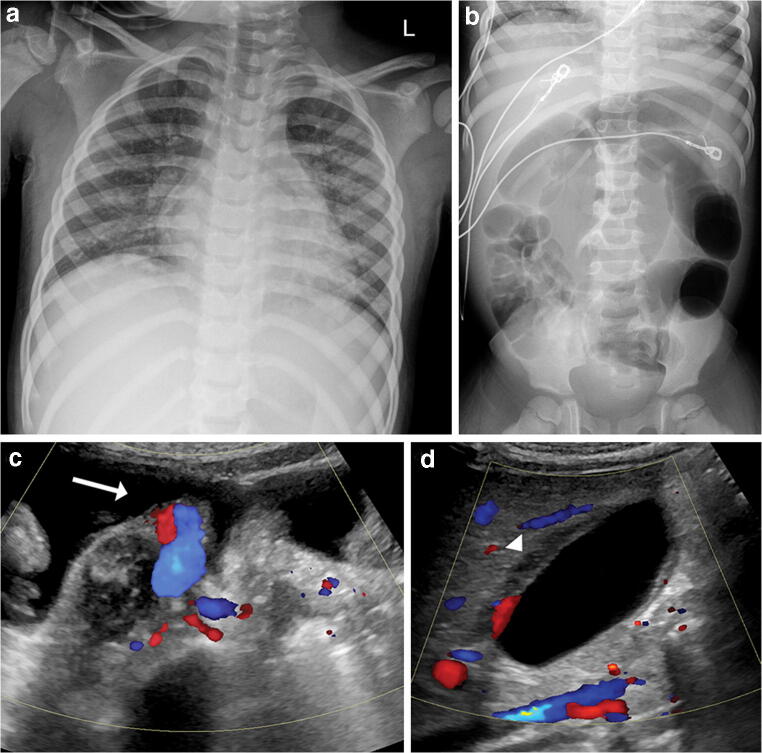
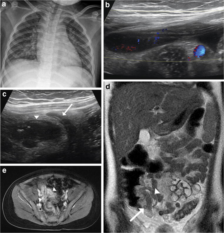
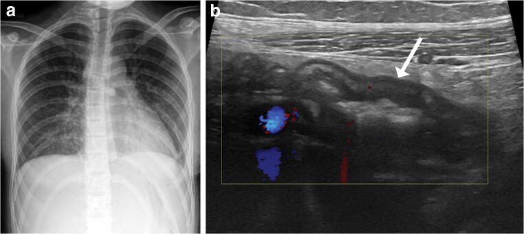
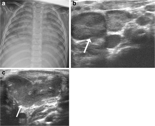
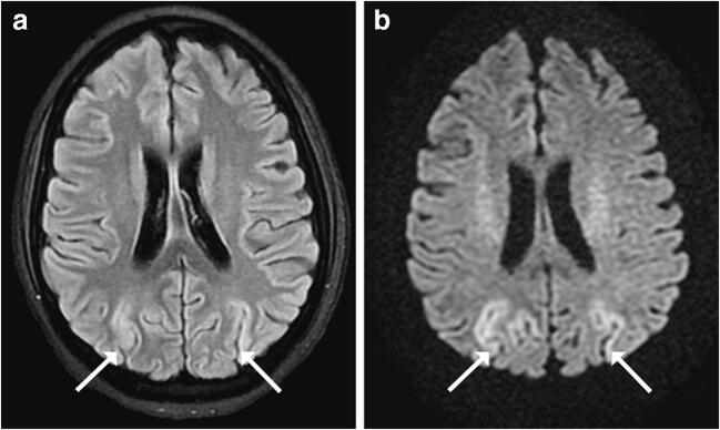
Results: Thirty-seven of 45 (82%) patients with chest radiographs had findings, with pulmonary opacities being the most common finding (n=27, 60%), most often bilateral and diffuse, followed by peribronchial thickening (n=26, 58%). Eight patients had normal chest radiographs. On abdominal imaging, small-volume ascites was the most common finding (n=7, 54%). Other findings included right lower quadrant bowel wall thickening (n=3, 23%), gallbladder wall thickening (n=3, 23%), and cervical (n=2) or abdominal (n=2) lymphadenopathy. Of the four patients with brain MRI, one had bilateral parieto-occipital abnormalities and another papilledema.

Conclusion: The diagnosis of MIS-C and its distinction from other pathologies should be primarily based on clinical presentation and laboratory evidence of inflammation because imaging findings are nonspecific. However, it should be considered in the setting of bilateral diffuse pulmonary opacities, peribronchial thickening, right lower quadrant bowel inflammation or unexplained ascites in a child presenting with Kawasaki-like symptoms and a history of COVID-19 infection or recent COVID-19 exposure.

Keywords: Abdomen; Children; Coronavirus disease 2019; Lungs; Magnetic resonance imaging; Multisystem inflammatory syndrome in children; Radiography; Ultrasound.


Conflict of interest statement

None

Figures

Fig. 1
Fig. 1
Imaging in a 4-year-old boy with a medical history of prematurity and mild asthma who presented with fever, abdominal pain, diarrhea, hypotensive shock and evidence of myocardial dysfunction with positive coronavirus disease 2019 (COVID-19) reverse transcription polymerase chain reaction (RT-PCR) and serology results. a Anteroposterior chest radiograph demonstrates bilateral perihilar opacities and peribronchial thickening. b Supine radiograph of the abdomen demonstrates abdominal distention with gaseous distention of the colon down to the rectum, suggesting colonic ileus. c Transverse US image of the right lower abdominal quadrant at the level of the iliac vasculature demonstrates small-volume ascites (arrow). d Sagittal sonographic image of the gallbladder demonstrates gallbladder wall thickening (arrowhead) but no gallstones
Fig. 2
Fig. 2
Imaging in a 7-year-old girl who presented with fever, abdominal pain, diarrhea, rash and evidence of myocardial dysfunction with a positive coronavirus disease 2019 (COVID-19) reverse transcription polymerase chain reaction (RT-PCR) result. This child was initially worked up for appendicitis. a Anteroposterior chest radiograph demonstrates bilateral perihilar opacities and peribronchial thickening. b, c Transverse US color Doppler (b) and gray-scale (c) images of the right lower abdominal quadrant demonstrate small-volume ascites and mesenteric edema adjacent to a borderline enlarged appendix (arrow), and a thickened loop of distal ileum (arrowhead). d, e Abdominal MRI coronal T2-W (d) and axial T1-W (e) fat-saturated post-gadolinium liver acquisition with volume acceleration (LAVA) images demonstrate a normal appendix (arrow) and terminal ileal and rectosigmoid wall thickening and hyperenhancement consistent with inflammation (arrowheads)
Fig. 3
Fig. 3
Imaging in a 15-year-old girl presenting with fever, abdominal pain, conjunctivitis, headaches and hypotensive shock with positive coronavirus disease 2019 (COVID-19) reverse transcription polymerase chain reaction (RT-PCR) and serology results. a Anteroposterior chest radiograph demonstrates bilateral perihilar opacities and peribronchial thickening. b Sagittal Doppler US image of the right lower abdominal quadrant demonstrates bowel wall thickening of the distal ileum (arrow) with surrounding mesenteric edema
Fig. 4
Fig. 4
Imaging in a 15-month-old girl who presented with fever, cervical lymphadenopathy, rash, hand and foot swelling with skin peeling, and evidence of myocardial dysfunction. She had a negative coronavirus disease 2019 (COVID-19) reverse transcription polymerase chain reaction (RT-PCR) result but had sick family contacts who had tested positive for COVID-19. a Anteroposterior chest radiograph demonstrates low lung volumes and diffuse airspace opacities. b, c Targeted transverse US images of the left lateral neck demonstrate cervical lymphadenopathy (arrows)
Fig. 5
Fig. 5
Brain imaging in a 14-year-old girl who initially presented with typical symptoms of multisystem inflammatory syndrome in children (MIS-C) including fever, abdominal pain and evidence of myocardial dysfunction with a positive coronavirus disease 2019 (COVID-19) serology result. She then developed thrombotic microangiopathy with hemolytic anemia and fluctuating mental status during admission. a, b Axial fluid-attenuated inversion recovery (FLAIR) MRI (a) and corresponding axial diffusion-weighted image (b) through the level of the bodies of the lateral ventricles and corona radiata demonstrate FLAIR hyperintensity/restricted diffusion involving the bilateral parieto-occipital cortices with mild cortical thickening (arrows)

Comment in

Similar articles

Cited by

References

    1. Riphagen S, Gomez X, Gonzalez-Martinez C, et al. Hyperinflammatory shock in children during COVID-19 pandemic. Lancet. 2020;395:1607–1608. doi: 10.1016/S0140-6736(20)31094-1. - DOI - PMC - PubMed
    1. CDC Health Alert Network (2020) Multisystem inflammatory syndrome in children (MIS-C) associated with coronavirus disease 2019 (COVID-19). Centers for Disease Control and Prevention website. https://emergency.cdc.gov/han/2020/han00432.asp. Accessed 10 June 2020
    1. Royal College of Paediatrics and Child Health – Health Policy Team (2020) Guidance: paediatric multisystem inflammatory syndrome temporally associated with COVID-19 (PIMS). Royal College of Paediatrics and Child Health website. https://www.rcpch.ac.uk/resources/guidance-paediatric-multisystem-inflam.... Accessed 10 June 2020
    1. Whittaker E, Bamford A, Kenny J, et al. Clinical characteristics of 58 children with a pediatric inflammatory multisystem syndrome temporally associated with SARS-CoV-2. JAMA. 2020;324:259–269. doi: 10.1001/jama.2020.10369. - DOI - PMC - PubMed
    1. Capone C, Subramony A, Sweberg T, et al. Characteristics, cardiac involvement, and outcomes of multisystem inflammatory syndrome of childhood associated with severe acute respiratory syndrome coronavirus 2 infection. J Pediatr. 2020;224:141–145. doi: 10.1016/j.jpeds.2020.06.044. - DOI - PMC - PubMed

Supplementary concepts