. 2021 Apr;41(4):1203-1221.
doi: 10.1007/s10792-020-01674-0. Epub 2021 Jan 12.

Degenerative lamellar macular holes: tractional development and morphological alterations

Affiliations

Degenerative lamellar macular holes: tractional development and morphological alterations

Andreas Bringmann et al. Int Ophthalmol. 2021 Apr.

Abstract

Purpose: The development of degenerative lamellar macular holes (DLH) is largely unclear. This study was aimed at documenting with spectral-domain optical coherence tomography the tractional development and morphological alterations of DLH.

Methods: A retrospective case series of 44 eyes of 44 patients is described.

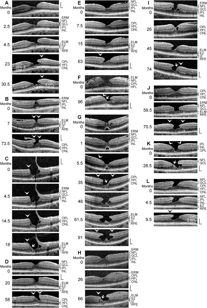
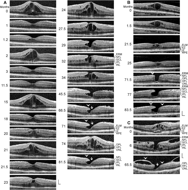
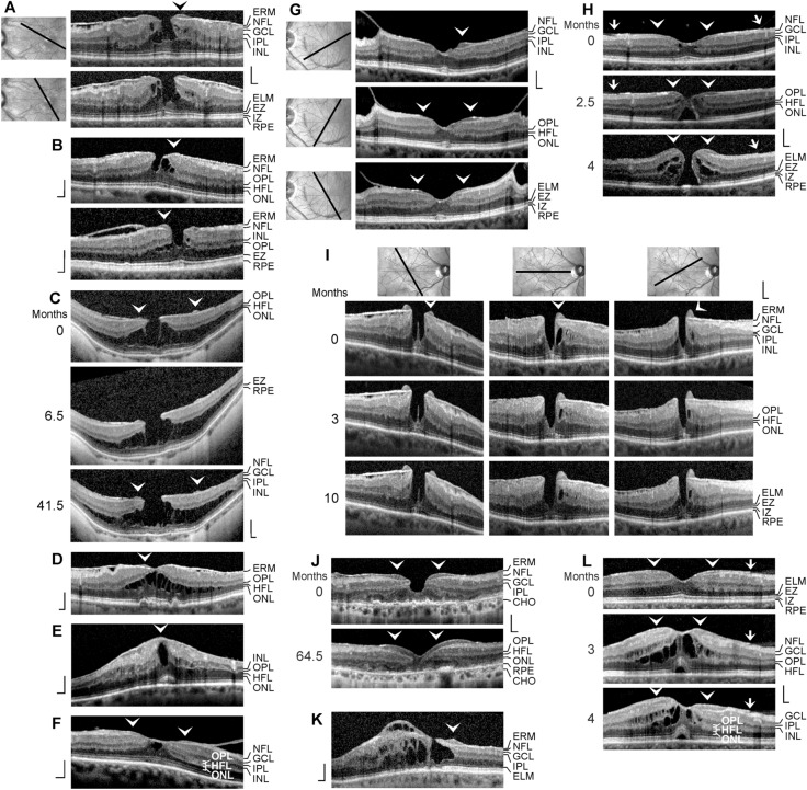
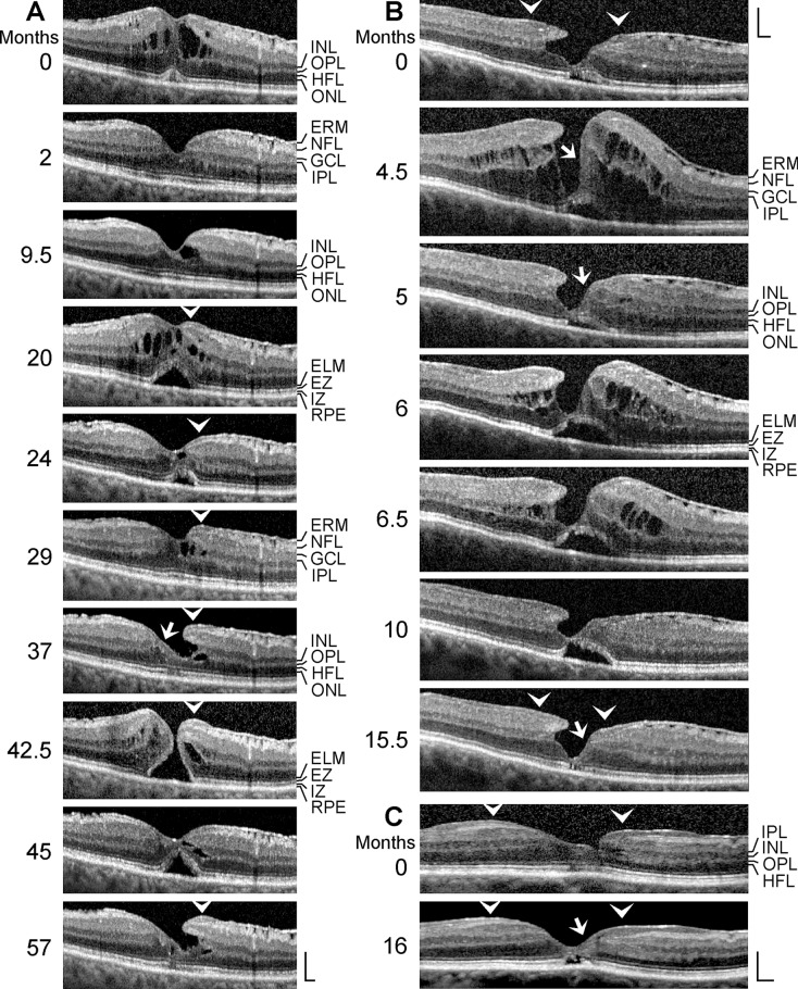
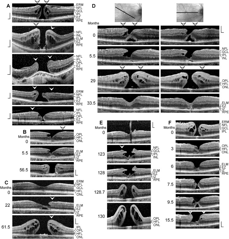
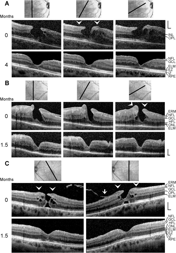
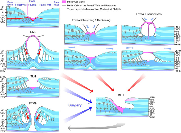
Results: The development of DLH is preceded for months or years by tractional deformations of the fovea due to the action of contractile epiretinal membranes (ERM) and/or the partially detached posterior hyaloid, or by cystoid macular edema (CME). DLH may develop after a tractional stretching and thickening of the foveal center, from a foveal pseudocyst, after a detachment of the foveola from the retinal pigment epithelium, a disruption of the foveal structure due to CME, and after surgical treatment of tractional lamellar or full-thickness macular holes (FTMH). The foveal configuration of a DLH can be spontaneously reestablished after short transient episodes of CME and a small FTMH. A DLH can evolve to a FTMH by traction of an ERM. Surgical treatment of a DLH may result in an irregular regeneration of the foveal center without photoreceptors.

Conclusions: Tractional forces play an important role in the development of DLH and in the further evolution to FTMH. It is suggested that a DLH is the result of a retinal wound repair process after a tractional disruption of the Müller cell cone and a degeneration of Henle fibers, to prevent a further increase in the degenerative cavitations.

Keywords: Fovea; Full-thickness macular hole; Lamellar macular hole; Müller cell cone; Müller glia.


Figures

Fig. 1
Fig. 1
Tractional development of degenerative lamellar holes (DLH). The images show SD-OCT scans through the fovea and parafovea of 12 eyes of 12 patients. The months after the first visit (0) are indicated left of the images. The arrowheads indicate lamellar macular hole-associated epiretinal proliferation (LHEP). The arrows indicate morphological connections between Müller cells in the foveola and LHEP. i Development of a DLH after surgical treatment of a tractional lamellar hole in an eye with macular pucker. Vitrectomy with internal limiting membrane and epiretinal membrane (ERM) peeling was performed 2.5 months after the first visit. Scale bars, 200 µm. ELM, external limiting membrane; ERM, epiretinal membrane; EZ, ellipsoid zone; GCL, ganglion cell layer; INL, inner nuclear layer; HFL, Henle fiber layer; IPL, inner plexiform layer; IZ, interdigitation zone; NFL, nerve fiber layer; ONL, outer nuclear layer; OPL, outer plexiform layer; RPE, retinal pigment epithelium
Fig. 2
Fig. 2
Cystoid macular edema may precede the development of a degenerative lamellar macular hole. ac The images show SD-OCT scans through the fovea and parafovea of 3 eyes of 3 patients. The months after the first visit (0) are indicated left of the images. The arrowheads indicate lamellar macular hole-associated epiretinal proliferation. The arrows indicate morphological connections between Müller cells in the foveola and the edges of nonelevated foveal walls. Scale bars, 200 µm. ELM, external limiting membrane; ERM, epiretinal membrane; EZ, ellipsoid zone; GCL, ganglion cell layer; HFL, Henle fiber layer; INL, inner nuclear layer; IPL, inner plexiform layer; IZ, interdigitation zone; NFL, nerve fiber layer; ONL, outer nuclear layer; OPL, outer plexiform layer; RPE, retinal pigment epithelium
Fig. 3
Fig. 3
Lamellar macular hole-associated epiretinal proliferation (LHEP) in various types of foveal defects. The images show SD-OCT scans through the fovea and parafovea of 13 eyes of 13 patients. The months after the first visit (0) are indicated left of the images. The orientations of the scans are shown above or left of the images. The arrowheads indicate LHEP. a, b Macular pseudoholes (MPH) with cleaved edges. c A tractional lamellar hole (TLH) with LHEP in an eye with high myopia. df Foveal pseudocysts. g Vitreomacular traction. h Development of a full-thickness macular hole by tangential traction exerted by the partially detached posterior hyaloid which adhered at the perifovea (arrows). The hole formation is associated with the formation of edematous cysts in the foveal walls. i, j Mixed types of a MPH and degenerative lamellar hole (DLH). k A DLH with cystoid macular edema (CME) in one foveal wall. Note that the ganglion cell layer (GCL) of this wall also contains edematous cysts. l Development of a CME from a fovea with LHEP. The arrow indicates a connection between the partially detached posterior hyaloid and an epiretinal membrane (ERM). Scale bars, 200 µm. CHO, choroidea; ELM, external limiting membrane; EZ, ellipsoid zone; HFL, Henle fiber layer; INL, inner nuclear layer; IPL, inner plexiform layer; IZ, interdigitation zone; NFL, nerve fiber layer; ONL, outer nuclear layer; OPL, outer plexiform layer; RPE, retinal pigment epithelium
Fig. 4
Fig. 4
Morphological alterations of degenerative lamellar holes. a–c The images show SD-OCT scans through the fovea and parafovea of 3 eyes of 3 patients. The months after the first visit (0) are indicated left of the images. The arrowheads indicate lamellar macular hole-associated epiretinal proliferation (LHEP). The arrows indicate morphological connections between Müller cells in the foveola and LHEP. Scale bars, 200 µm. ELM, external limiting membrane; ERM, epiretinal membrane; EZ, ellipsoid zone; GCL, ganglion cell layer; HFL, Henle fiber layer; INL, inner nuclear layer; IPL, inner plexiform layer; IZ, interdigitation zone; NFL, nerve fiber layer; ONL, outer nuclear layer; OPL, outer plexiform layer; RPE, retinal pigment epithelium
Fig. 5
Fig. 5
Degenerative lamellar holes (DLH) and full-thickness macular holes (FTMH). The images show SD-OCT scans through the fovea and parafovea of 10 eyes of 10 patients. The months after the first visit (0) are indicated left of the images. In c, the orientations of the scans are indicated above the images. The arrowheads indicate lamellar macular hole-associated epiretinal proliferation (LHEP). The arrows indicate morphological connections between Müller cells in the foveola and LHEP. a Five eyes with a FTMH with LHEP. b–d Development of a DLH into a FTMH. e Tractional development of a DLH and development into a FTMH. Vitrectomy with ILM and ERM peeling was performed 31 (d) and 128.2 months (e) after the first visit, respectively. f Development of a DLH after surgical closure of a FTMH. Vitrectomy with ILM and ERM peeling was performed 1.5 months after the first visit. Scale bars, 200 µm. ELM, external limiting membrane; EZ, ellipsoid zone; GCL, ganglion cell layer; HFL, Henle fiber layer; INL, inner nuclear layer; IPL, inner plexiform layer; IZ, interdigitation zone; NFL, nerve fiber layer; ONL, outer nuclear layer; OPL, outer plexiform layer; RPE, retinal pigment epithelium
Fig. 6
Fig. 6
Foveal regeneration after surgical removal of lamellar macular hole-associated epiretinal proliferation (LHEP) by vitrectomy with internal limiting and epiretinal membrane (ERM) peeling. a–c. The images show SD-OCT scans through the fovea and parafovea of 3 eyes of 3 patients recorded before (above) and after surgery (below). The orientations of the scans are shown above the images. The months after the first visit (0) are indicated left of the images. The arrowheads indicate LHEP. The arrow indicates the adhesion of the partially detached posterior hyaloid to the parafoveal tissue. Surgery was performed 1.5 months (a), one month (b), and one day (c) after the first visit, respectively. Scale bars, 200 µm. ELM, external limiting membrane; EZ, ellipsoid zone; GCL, ganglion cell layer; HFL, Henle fiber layer; INL, inner nuclear layer; IZ, interdigitation zone; NFL, nerve fiber layer; ONL, outer nuclear layer; OPL, outer plexiform layer; RPE, retinal pigment epithelium
Fig. 7
Fig. 7
Schematic summary of different modes of the development of degenerative lamellar holes (DLH) induced by traction onto the fovea. The image above shows a schematic cross section through the normal fovea. The blue arrows indicate anterior hyaloidal traction and horizontal traction exerted by epiretinal membranes (ERM), respectively. The red arrows indicate anterior traction exerted by the stretched Müller cells of the foveal walls which causes a detachment of the central outer retina from the retinal pigment epithelium (RPE). The development of DLH may be preceded by cystoid macular edema (CME), which tractionally disrupts the foveal structure, or by traction onto the fovea exerted by the partially detached posterior hyaloid and/or ERM which causes a stretching and thickening of the foveola associated with or not a detachment of the central fovea from the RPE. The traction may also produce foveal pseudocysts. In addition, surgical treatment of tractional lamellar holes (TLH) or full-thickness macular holes (FTMH) may result in the development of DLH. A DLH may evolve into a FTMH. ELM, external limiting membrane; EZ, ellipsoid zone; GCL, ganglion cell layer; HFL, Henle fiber layer; INL, inner nuclear layer; IPL, inner plexiform layer; IZ, interdigitation zone; NFL, nerve fiber layer; ONL, outer nuclear layer; OPL, outer plexiform layer

Similar articles

Cited by

References

    1. Yamada E. Some structural features of the fovea centralis in the human retina. Arch Ophthalmol. 1969;82:151–159. - PubMed
    1. Gass JDM. Müller cell cone, an overlooked part of the anatomy of the fovea centralis: Hypotheses concerning its role in the pathogenesis of macular hole and foveomacualr retinoschisis. Arch Ophthalmol. 1999;117:821–823. - PubMed
    1. Syrbe S, Kuhrt H, Gärtner U, Habermann G, Wiedemann P, Bringmann A, Reichenbach A. Müller glial cells of the primate foveola: An electron microscopical study. Exp Eye Res. 2018;167:110–117. - PubMed
    1. Bringmann A, Syrbe S, Görner K, Kacza J, Francke M, Wiedemann P, Reichenbach A. The primate fovea: structure, function and development. Prog Retin Eye Res. 2018;66:49–84. - PubMed
    1. Bringmann A, Unterlauft JD, Wiedemann R, Rehak M, Wiedemann P. Morphology of partial-thickness macular defects: Presumed roles of Müller cells and tissue layer interfaces of low mechanical stability. Int J Retina Vitreous. 2020;6:28. - PMC - PubMed

LinkOut - more resources