Review
. 2021 Apr;10(7):e2001689.
doi: 10.1002/adhm.202001689. Epub 2021 Jan 12.

Effects of Mesenchymal Stem Cell-Derived Paracrine Signals and Their Delivery Strategies

Affiliations
Review

Effects of Mesenchymal Stem Cell-Derived Paracrine Signals and Their Delivery Strategies

Calvin Chang et al. Adv Healthc Mater. 2021 Apr.

Abstract

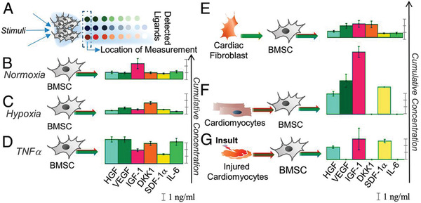
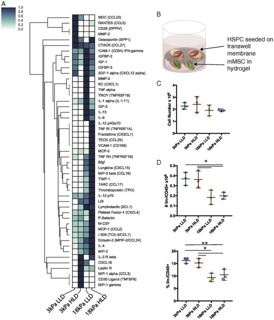
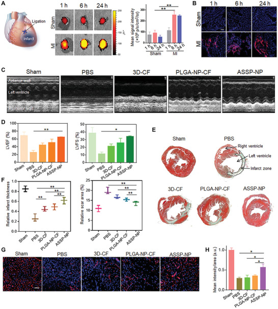
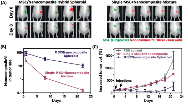
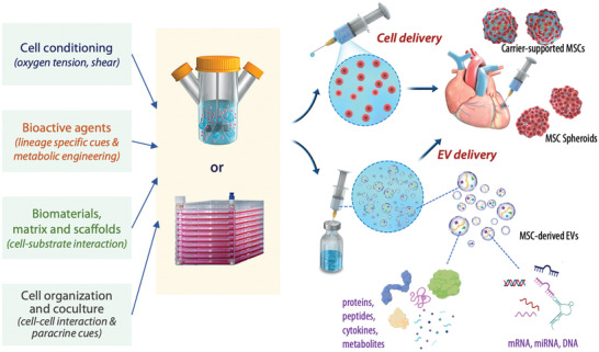
Mesenchymal stem cells (MSCs) have been widely studied as a versatile cell source for tissue regeneration and remodeling due to their potent bioactivity, which includes modulation of inflammation response, macrophage polarization toward proregenerative lineage, promotion of angiogenesis, and reduction in fibrosis. This review focuses on profiling the effects of paracrine signals of MSCs, commonly referred to as the secretome, and highlighting the various engineering approaches to tune the MSC secretome. Recent advances in biomaterials-based therapeutic strategies for delivery of MSCs and MSC-derived secretome in the form of extracellular vesicles are discussed, along with their advantages and challenges.

Keywords: biomaterials; extracellular vesicles; mesenchymal stem cells; microRNA; paracrine signaling; secretome.


Conflict of interest statement

The authors declare no conflict of interest.

Figures

Figure 1
Figure 1
Secretion of BMSCs in response to specific environmental signals.[ 84 ] A) Secretion profiles were measured using detector microspots in an antibody array separated from the cell culture chamber by pillars at the 12 h time point. Cells displayed different secretory profiles in B) normoxic conditions, C) hypoxia condition, D) TNF‐α stimulation, E) conditioned media from cardiac fibroblasts, F) conditioned media from human induced pluripotent stem cell‐derived cardiomyocytes (hiPSC‐CM), and G) conditioned media with hiPSC‐CM insulted with peroxide to mimic ischemia reperfusion injury. Reproduced with permission.[ 84 ] Copyright 2019, National Academy of Sciences.
Figure 2
Figure 2
mMSCs‐secreted cytokines in response to substrate stiffness.[ 91 ] A) Cytokine antibody array analysis was performed on conditioned media from mMSCs cultured in alginate hydrogels of varying stiffnesses and adhesion ligand (Arg‐Gly‐Asp) densities for 2–3 days, with values normalized to internal positive control and maximum signal for each material. B) Schematic of MSC and hematopoietic stem and progenitor cell (hSPC) coculture system. C) Viable cell number measured by flow cytometry after 1 week of coculture and D) number and percentage of CD45+/lin cells from transwell membrane measured by flow cytometry after 1 week of coculture. Reproduced with permission.[ 91 ] Copyright 2018, National Academy of Sciences.
Figure 3
Figure 3
Intravenous administration of artificial stem cell spheroid nanoparticles (ASSP‐NP) in a mouse MI model for cardiac repair.[ 112 ] A) Ex vivo fluorescent imaging of mouse hearts and quantitative analysis of fluorescence intensities at different time points after tail vein injection of ASSP‐NPs in sham and MI mice. B) ASSP‐NP distribution in sham (nonischemic) and MI (ischemic) cardiac tissue at different time points following intravenous injection. C) Echocardiography images of left ventricular wall motion with or without treatments following MI surgery at 28 days using PBS, cocktail factor containing conditioned media from 3D SSP (3D‐CF), PLGA nanoparticles (PLGA‐NP‐CF), and targeted ASSP nanoparticles (ASSP‐NP) with red blood cell membrane and platelet membrane coatings. D) Quantification of echocardiography functional assay, including left ventricular ejection fraction (LVEF) percentage and left ventricular fractional shortening (LVFS) percentage. E) Masson's trichrome staining of midpapillary sections of the heart at 28 days after MI. F) Quantitative analysis of infarct wall thickness and scar area. G) Representative images and H) image‐based quantification of blood vessels in cardiac ischemic conditions by anti‐CD31 antibody immunostaining at 28 days after treatments. Reproduced with permission.[ 112 ] Copyright 2020, American Association for the Advancement of Science.
Figure 4
Figure 4
MSC/nanocomposite spheroid improves tumor homing of MSCs and reduction tumor volume growth.[ 113 ] A) Mice treated with hybrid spheroids generated with a microfluidics‐based approach and PEGylated DNA template or single MSC‐nanocomposite mixture through injection at the boundary of the tumor site on days 0 and 2, and quantification B,C) of tumor homing and inhibition. Reproduced with permission.[ 113 ] Copyright 2019, American Chemical Society.
Figure 5
Figure 5
EV Dose‐response data for suppression of neuroinflammation after traumatic brain injury (TBI).[ 115 ] A) Immunohistochemistry staining of brain sections after TBI mouse model. B) Decreased levels of IL‐1β in a dose‐dependent manner of PBS or EVs administrated 1 h after TBI measured by ELISA on homogenates from ipsilateral brain sections isolated 12 h after TBI. Reproduced with permission.[ 115 ] Copyright 2015, National Academy of Sciences.
Figure 6
Figure 6
Tuning of the MSC secretome profile for the delivery of engineered MSCs and paracrine cues. MSC secretome can be modulated by adjusting cell culture conditions, supplementing bioactive agents, employing biomaterials matrix or scaffold, and modulating cell–cell interactions. These engineering approaches can be adopted to adjust the quantities and components of MSC paracrine signals prior to cell delivery to the treatment site. For cell delivery, the engineered MSCs can be injected in the form of MSC spheroids or carrier‐supported MSCs, or biomaterials delivered with the MSCs. The secretome can also be delivered in the form of EVs enriched from the cultured MSCs prior to delivery. The EVs contain proteins, peptides, cytokines, metabolites, as well as nucleic acids including mRNA, miRNA, and DNA.

References

    1. Caplan A. I., J. Orthop. Res. 1991, 9, 641. - PubMed
    1. Dominici M., Blanc K. Le, Mueller I., Slaper‐Cortenbach I., Marini F. C., Krause D. S., Deans R. J., Keating A., Prockop D. J., Horwitz E. M., Cytotherapy 2006, 8, 315. - PubMed
    1. Zuk P. A., Zhu M. I. N., Mizuno H., Huang J., Futrell J. W., Katz A. J., Benhaim P., Lorenz H. P., Hedrick M. H., Tissue Eng. 2001, 7, 211. - PubMed
    1. Noronha Nc N. D. C., Mizukami A., Caliári‐Oliveira C., Cominal J. G., Rocha J. L. M., Covas D. T., Swiech K., Malmegrim K. C. R., Stem Cell Res. Ther. 2019, 10, 131. - PMC - PubMed
    1. Shi S., Gronthos S., J. Bone Miner. Res. 2003, 18, 696. - PubMed

Publication types

LinkOut - more resources