Spinal epidural lipomatosis: a rare association of Cushing's disease
- PMID: 33434165
- PMCID: PMC7576635
- DOI: 10.1530/EDM-20-0111
Spinal epidural lipomatosis: a rare association of Cushing's disease
Abstract
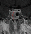
Summary: Excess cortisol is associated with hypertrophy and redistribution of adipose tissue leading to central obesity which is classically seen in Cushing's syndrome. Abnormal accumulation of fatty tissue in the spinal canal is most commonly associated with chronic steroid therapy and rarely reported with endogenous Cushing's syndrome. Herein, we describe a case of spinal epidural lipomatosis (SEL) associated with Cushing's disease. A 17-year-old man was referred with lower limb weakness, weight gain, multiple stretch marks, back pain and loss of height. He had clinical and biochemical features of Cushing's syndrome. MRI and Inferior Petrosal Sinus Sampling (IPSS) confirmed a pituitary adenoma as the source. On day 1 post trans-sphenoidal adenectomy he developed spastic paraparesis with a sensory deficit to the level of T5. MRI spine showed increased fat deposition in the spinal canal from T2 to T9 consistent with a diagnosis of SEL. He was managed conservatively and made a good recovery following restoration of eucortisolism and a period of rehabilitation.
Learning points: SEL is a serious complication of glucocorticoid excess and should be considered in any patient presenting with new lower limb neurological symptoms associated with hypercortisolism. It is important to distinguish symptomatic SEL from cortisol-induced proximal myopathy by good history and clinical examination. MRI of the spine is the gold standard investigation for making a diagnosis of SEL. Restoration of eucortisolism can lead to resolution of fat accumulation and good neurological outcome.
Figures


References
LinkOut - more resources
Full Text Sources

