

Using stable isotope labeling to advance our understanding of Alzheimer's disease etiology and pathology
- PMID: 33434345
- PMCID: PMC8273190
- DOI: 10.1111/jnc.15298
Using stable isotope labeling to advance our understanding of Alzheimer's disease etiology and pathology
Abstract
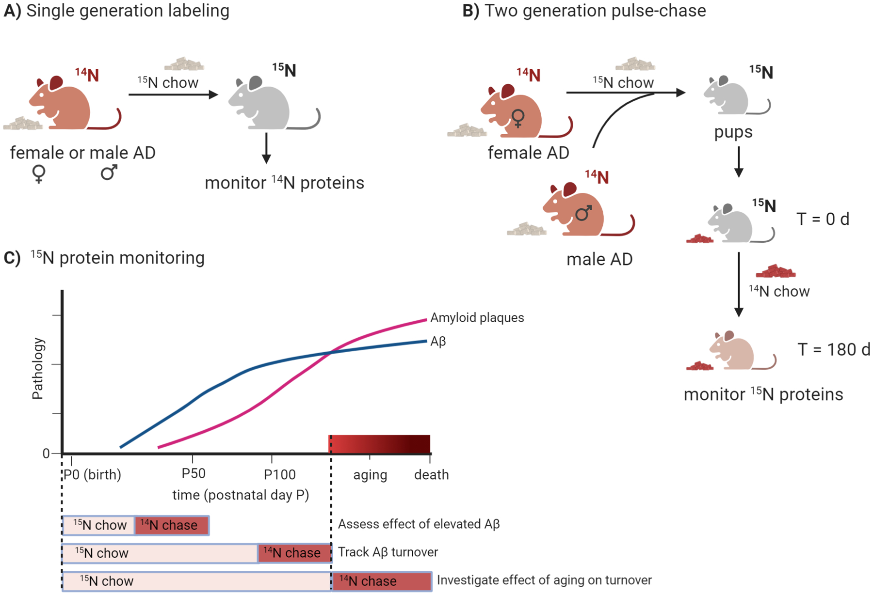
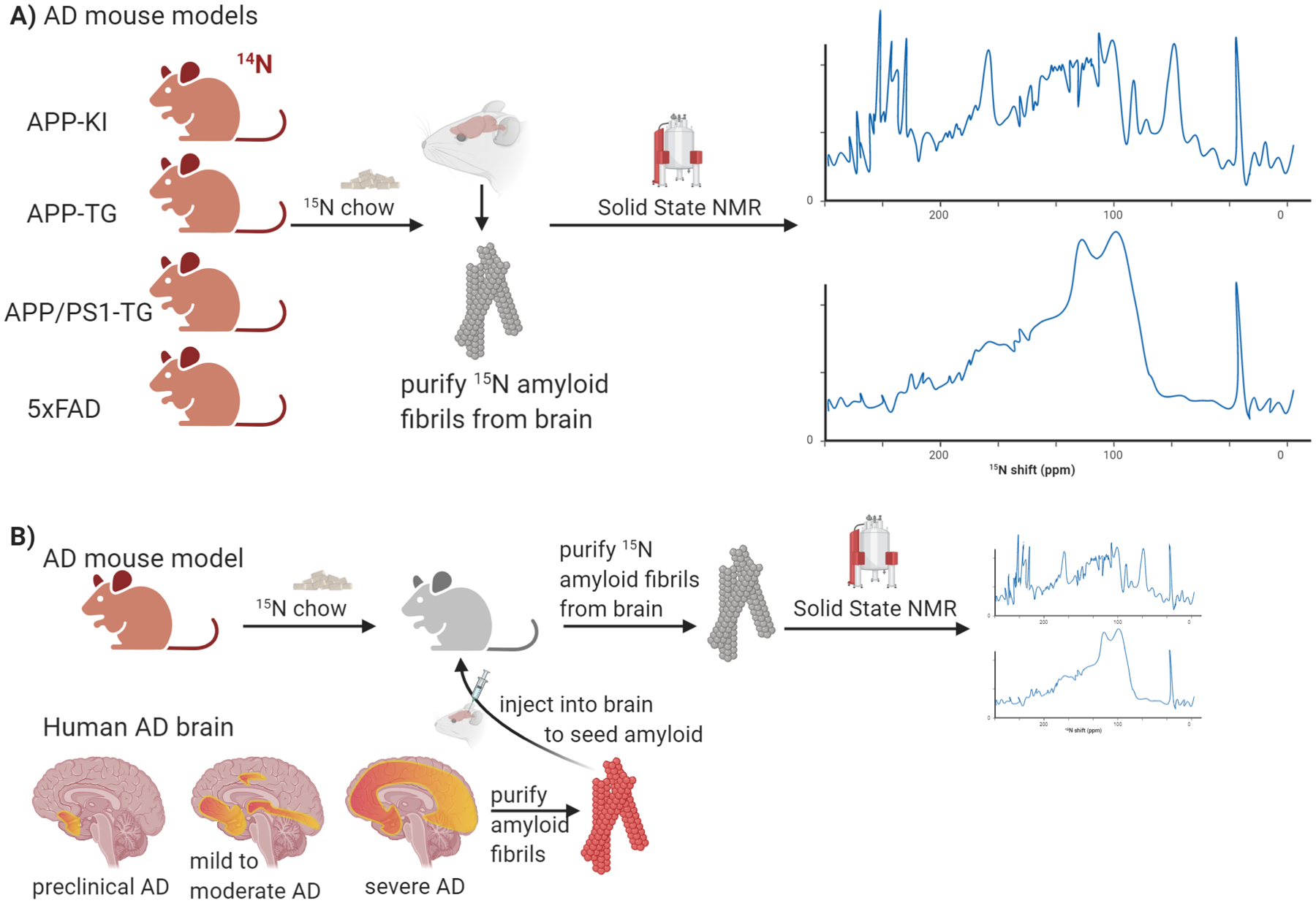
Stable isotope labeling with mass spectrometry (MS)-based proteomic analysis has become a powerful strategy to assess protein steady-state levels, protein turnover, and protein localization. Applying these analyses platforms to neurodegenerative disorders may uncover new aspects of the etiology of these devastating diseases. Recently, stable isotopes-MS has been used to investigate early pathological mechanisms of Alzheimer's disease (AD) with mouse models of AD-like pathology. In this review, we summarize these stable isotope-MS experimental designs and the recent application in the context of AD pathology. We also describe our current efforts aimed at using nuclear magnetic resonance (NMR) analysis of stable isotope-labeled amyloid fibrils from AD mouse model brains. Collectively, these methodologies offer new opportunities to study proteome changes in AD and other neurodegenerative diseases by elucidating mechanisms to target for treatment and prevention.
Keywords: APP Knock-In Mice; Alzheimer's disease; Amyloid-β; mass spectrometry; proteomics; stable isotopes.
© 2021 International Society for Neurochemistry.
Conflict of interest statement
Conflict of Interest
The authors declare no conflicts of interest.
Figures

Similar articles
-
Neuroproteomic tools for battling Alzheimer's disease.Proteomics. 2016 Nov;16(22):2847-2853. doi: 10.1002/pmic.201600211. Epub 2016 Oct 14. Proteomics. 2016. PMID: 27633846 Review.
-
Capillary Electrophoresis-High Resolution Mass Spectrometry for Measuring In Vivo Arginine Isotope Incorporation in Alzheimer's Disease Mouse Models.J Am Soc Mass Spectrom. 2021 Jun 2;32(6):1448-1458. doi: 10.1021/jasms.1c00055. Epub 2021 May 24. J Am Soc Mass Spectrom. 2021. PMID: 34028275
-
Development of isotope labeling liquid chromatography mass spectrometry for mouse urine metabolomics: quantitative metabolomic study of transgenic mice related to Alzheimer's disease.J Proteome Res. 2014 Oct 3;13(10):4457-69. doi: 10.1021/pr500828v. Epub 2014 Sep 15. J Proteome Res. 2014. PMID: 25164377
-
Early Presymptomatic Changes in the Proteome of Mitochondria-Associated Membrane in the APP/PS1 Mouse Model of Alzheimer's Disease.Mol Neurobiol. 2018 Oct;55(10):7839-7857. doi: 10.1007/s12035-018-0955-6. Epub 2018 Feb 22. Mol Neurobiol. 2018. PMID: 29468564
-
Does HIV infection contribute to increased beta-amyloid synthesis and plaque formation leading to neurodegeneration and Alzheimer's disease?J Neurovirol. 2019 Oct;25(5):634-647. doi: 10.1007/s13365-019-00732-3. Epub 2019 Mar 13. J Neurovirol. 2019. PMID: 30868421 Review.
Cited by
-
Coupling Stable Isotope Labeling and Liquid Chromatography-Trapped Ion Mobility Spectrometry-Time-of-Flight-Tandem Mass Spectrometry for De Novo Mosquito Ovarian Lipid Studies.Anal Chem. 2022 Apr 26;94(16):6139-6145. doi: 10.1021/acs.analchem.1c05090. Epub 2022 Apr 14. Anal Chem. 2022. PMID: 35420029 Free PMC article.
-
Mass Spectrometry Imaging in Alzheimer's Disease.Brain Connect. 2023 Aug;13(6):319-333. doi: 10.1089/brain.2022.0057. Epub 2023 Apr 24. Brain Connect. 2023. PMID: 36905365 Free PMC article.
-
Determining and interpreting protein lifetimes in mammalian tissues.Trends Biochem Sci. 2023 Feb;48(2):106-118. doi: 10.1016/j.tibs.2022.08.011. Epub 2022 Sep 23. Trends Biochem Sci. 2023. PMID: 36163144 Free PMC article. Review.
-
Lipidomics and proteomics: An integrative approach for early diagnosis of dementia and Alzheimer's disease.Front Genet. 2023 Feb 9;14:1057068. doi: 10.3389/fgene.2023.1057068. eCollection 2023. Front Genet. 2023. PMID: 36845373 Free PMC article. Review.
-
The cochlea is built to last a lifetime.Hear Res. 2023 Sep 1;436:108821. doi: 10.1016/j.heares.2023.108821. Epub 2023 Jun 1. Hear Res. 2023. PMID: 37295280 Free PMC article. Review.
References
-
- (2020). 2020 Alzheimer’s disease facts and figures. Alzheimers Dement. - PubMed
-
- Andersen JS, Lam YW, Leung AK, Ong SE, Lyon CE, Lamond AI, and Mann M (2005). Nucleolar proteome dynamics. Nature 433, 77–83. - PubMed
-
- Andrew RJ, Fisher K, Heesom KJ, Kellett KAB, and Hooper NM (2019). Quantitative interaction proteomics reveals differences in the interactomes of amyloid precursor protein isoforms. J Neurochem 149, 399–412. - PubMed
-
- Balch WE, Morimoto RI, Dillin A, and Kelly JW (2008). Adapting proteostasis for disease intervention. Science 319, 916–919. - PubMed
Publication types
MeSH terms
Substances
Grants and funding
LinkOut - more resources
Full Text Sources
Other Literature Sources
Medical
