. 2021 Mar:150:105258.
doi: 10.1016/j.nbd.2021.105258. Epub 2021 Jan 9.

Regulation of distinct caspase-8 functions in retinal ganglion cells and astroglia in experimental glaucoma

Affiliations

Regulation of distinct caspase-8 functions in retinal ganglion cells and astroglia in experimental glaucoma

Xiangjun Yang et al. Neurobiol Dis. 2021 Mar.

Abstract

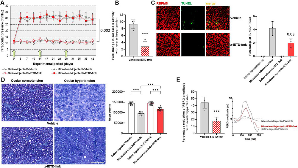
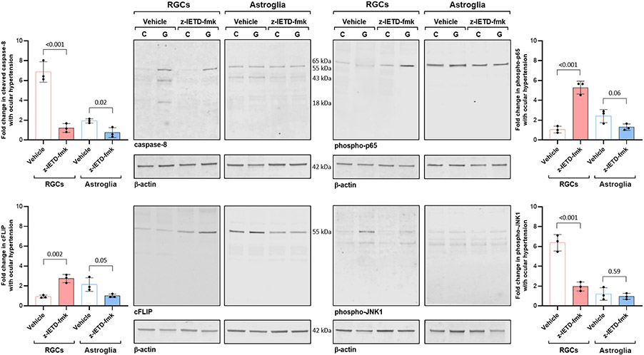
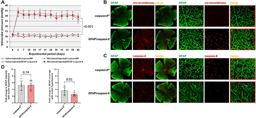
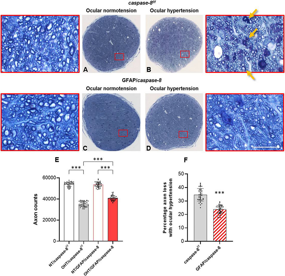
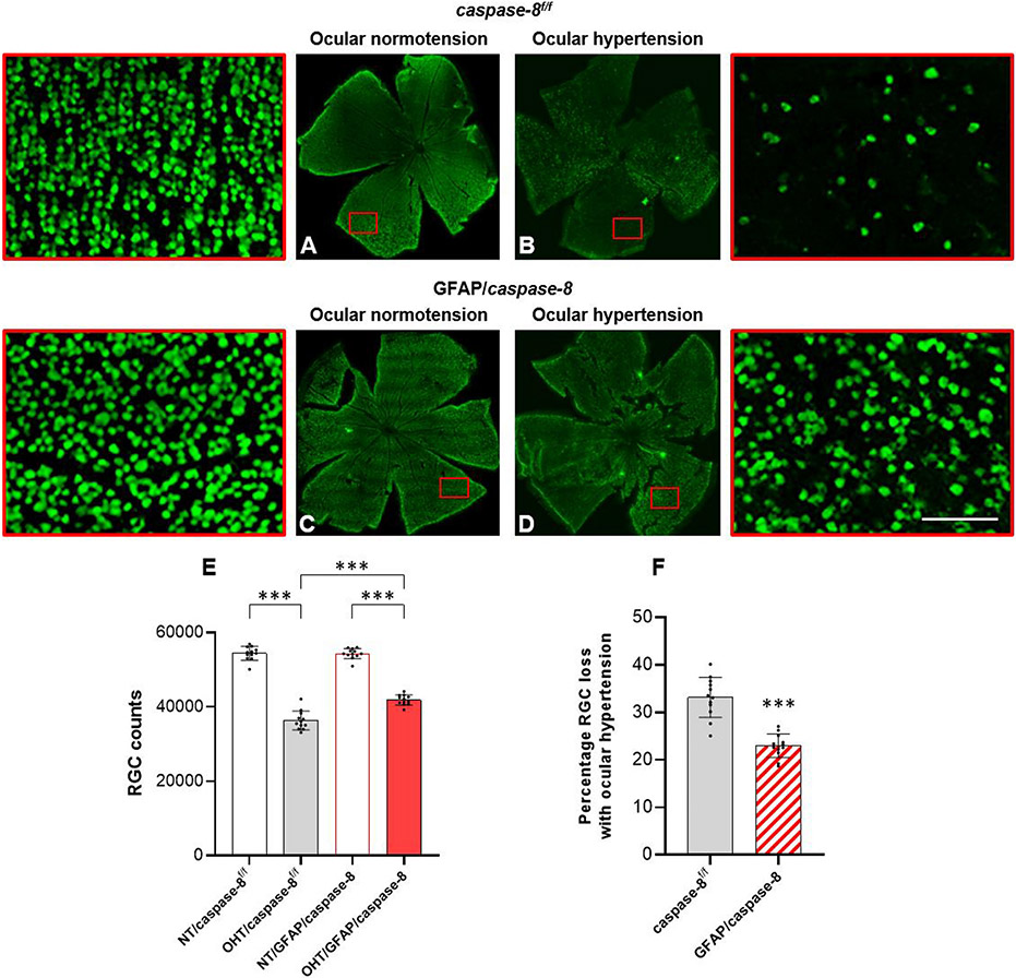
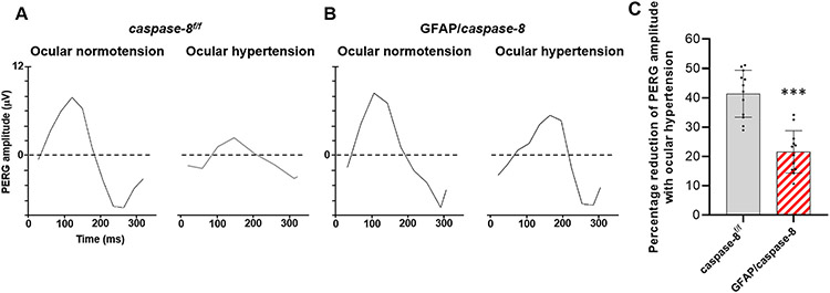
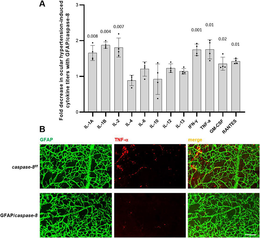
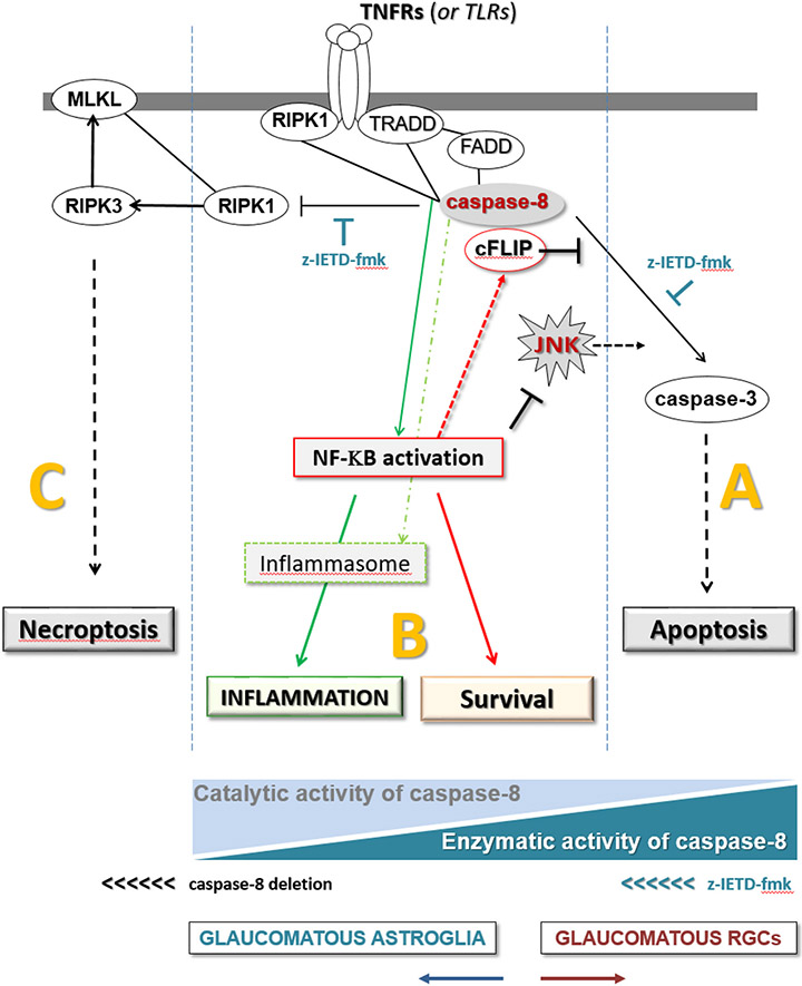
Retinal ganglion cells (RGCs) expanding from the retina to the brain are primary victims of neurodegeneration in glaucoma, a leading cause of blindness; however, the neighboring astroglia survive the glaucoma-related stress and promote neuroinflammation. In light of diverse functions of caspase-8 in apoptosis, cell survival, and inflammation, this study investigated the importance of caspase-8 in different fates of glaucomatous RGCs and astroglia using two experimental approaches in parallel. In the first approach, cell type-specific responses of RGCs and astroglia to a caspase-8 cleavage-inhibiting pharmacological treatment were studied in rat eyes with or without experimentally induced glaucoma. The second approach utilized an experimental model of glaucoma in mice in which astroglial caspase-8 was conditionally deleted by cre/lox. Findings of these experiments revealed cell type-specific distinct processes that regulate caspase-8 functions in experimental glaucoma, which are involved in inducing the apoptosis of RGCs and promoting the survival and inflammatory responses of astroglia. Deletion of caspase-8 in astroglia protected RGCs against glia-driven inflammatory injury, while the inhibition of caspase-8 cleavage inhibited apoptosis in RGCs themselves. Various caspase-8 functions impacting both RGC apoptosis and astroglia-driven neuroinflammation may suggest the multi-target potential of caspase-8 regulation to provide neuroprotection and immunomodulation in glaucoma.

Keywords: Astroglia; Caspase-8; Glaucoma; Neurodegeneration; Neuroinflammation.


Figures

Fig. 1.
Fig. 1.. Experimental modeling of ocular hypertension-induced glaucoma in rats and pharmacological inhibition of caspase-8 cleavage.
A. Intraocular pressure curves through the 6-weeks experimental period. Green arrows show the time points for z-IETD-fmk injections. Microbead injections resulted in a significant increase in intraocular pressure (P = 0.002) that did not change with z-IETD-fmk treatment or injections of vehicle only (P = 0.56; n > 40/group). B. Caspase-8 activity assay detected a significant decrease in z-IETD-fmk treated retinas (***P < 0.001; n > 4/group). C. TUNEL labeling (green) in retinal whole mounts showed a significant decrease (P = 0.03) in the number RBPMS-labeled RGCs (red) in z-IETD-fmk-injected ocular hypertensive eyes than vehicle-injected ocular hypertensive controls (n > 4/group; scale bar, 100 μm). D. Axon counts in optic nerve cross-sections indicated approximately 40% protection against ocular hypertension-induced axon loss with z-IETD-fmk treatment (***P < 0.001; n > 36/group; scale bar, 10 μm). E. The PERG amplitude was also preserved in z-IETD-fmk-treated ocular hypertensive eyes compared to vehicle-injected ocular hypertensive controls (n > 4/group). Data are presented as mean ± SD, P values were obtained using a one-way ANOVA.
Fig. 2.
Fig. 2.. RGC and astroglia responses to z-IETD-fmk treatment.
RGC and astroglia proteins were isolated from rat eyes with experimental glaucoma (G) or normotensive controls (C) that were injected with z-IETD-fmk or vehicle only. Immunoblots were probed with antibodies to caspase-8, phospho-p65, cFLIP, or phospho-JNK1 (n > 3/group). Data are presented as mean ± SD, P values (obtained using a one-way ANOVA) for the statistical comparison of experimental groups are given in each graph.
Fig. 3.
Fig. 3.. Experimental modeling of ocular hypertension-induced glaucoma in mice and cre/lox-based deletion of astroglial caspase-8.
A. Intraocular pressure curves through the 6-weeks experimental period. Microbead injections resulted in a significant increase in intraocular pressure (***P < 0.001) that was similar in GFAP/caspase-8 mice and caspase-8f/f controls (P = 0.20; n > 39/group). B. Immunolabeling of retinal whole mounts demonstrated cre-recombinase (red) expression in GFAP+ astroglia (green) after tamoxifen-injection in GFAP/caspase-8 mice. C. Based on immunolabeling of retinal whole mounts, no caspase-8 (red) expression was detectable in GFAP+ astroglia (green) in GFAP/caspase-8 mice. Red boxed areas are shown in higher magnification (scale bar, 100 μm). D. Image analysis detected no alteration in the intensity of GFAP immunolabeling (reflecting the GFAP expression level of astroglia) with caspase-8 deletion. However, the coverage of GFAP immunolabeling (reflecting the size and density of individual cells) was significantly lower in GFAP/caspase-8 retinas compared with caspase-8f/f controls (n > 20/group). Data are presented as mean ± SD, P values (obtained using a one-way ANOVA) for the statistical comparison of experimental groups are given in each graph.
Fig. 4.
Fig. 4.. Effects of GFAP/caspase-8 on optic nerve axon counts.
Presented are composite images of optic nerve cross-sections stained with 2% toluidine blue. Red boxed areas are shown in higher magnification (scale bar, 10 μm). Compared to normotensive caspase-8f/f control mice (A), there was a prominent axon loss and gliosis in ocular hypertensive eyes of caspase-8f/f controls (B). No alteration in optic nerve structure was detectable in normotensive mice with GFAP-targeting caspase-8 deletion (C). However, compared to ocular hypertensive caspase-8f/f controls, optic nerve structure was well preserved in ocular hypertensive eyes of GFAP/caspase-8 mice (D). E. The number of remaining axons was significantly higher in ocular hypertensive GFAP/caspase-8 mice than ocular hypertensive caspase-8f/f controls. F. Astroglial caspase deletion provided >30% protection against axon loss in experimental glaucoma. Yellow arrows show degenerating axons and myelin debris. Data (mean ± SD) represents 39 mouse eyes per group (***one-way ANOVA, P < 0.001).
Fig. 5.
Fig. 5.. Effects of GFAP/caspase-8 on RGC soma counts.
Presented are composite images of the whole mounted retinas immunolabeled for RBPMS, an RGC marker. Red boxed areas are shown in higher magnification (scale bar, 100 μm). Compared to normotensive caspase-8f/f control mice (A), there was a visible loss of RBPMS-labeled RGC somas (green) in ocular hypertensive eyes of caspase-8f/f controls (B). No alteration in RBPMS labeling was detectable in normotensive mice with GFAP-targeting caspase-8 deletion (C). However, compared to ocular hypertensive caspase-8f/f controls, RBPMS-labeled RGCs were well protected in ocular hypertensive eyes of GFAP/caspase-8 mice (D). E. The number of RBPMS-labeled RGCs was significantly higher in ocular hypertensive GFAP/caspase-8 mice than ocular hypertensive caspase-8f/f controls. F. Astroglial caspase deletion provided an approximately 30% protection against RGC soma loss in experimental glaucoma. Data (mean ± SD) represents 12 mouse eyes per group (***one-way ANOVA, P < 0.001).
Fig. 6.
Fig. 6.. Effects of GFAP/caspase-8 on pattern electroretinography (PERG) responses.
A. Shown are PERG responses in GFAP/caspase-8 mice and caspase-8f/f controls with or without experimentally induced ocular hypertension (A). The bar graphs show PERG amplitude in each group (B). Compared to normotensive eyes, ocular hypertensive eyes showed a reduction in PERG amplitude. However, deletion of astroglial caspase-8 resulted in preserved PERG amplitude in ocular hypertensive eyes. Data (mean ± SD) represents 12 mouse eyes per group (***one-way ANOVA, P < 0.001).
Fig. 7.
Fig. 7.. Effects of GFAP/caspase-8 on pro-inflammatory cytokine production.
A. The bar graph presents the fold decrease in ocular hypertension-induced cytokine titers with GFAP/caspase-8 relative to caspase-8f/f (statistical significance are shown by P values obtained using a one-way ANOVA). Data (mean ± SD) were obtained using a minimum of three new samples per group. B. Immunolabeling of retinal whole mounts supported decreased cytokine response (TNF-α, red) of GFAP-labeled astroglia (green) to ocular hypertension in GFAP/caspase-8 mice compared to caspase-8f/f ocular hypertensive controls (scale bar, 100 μm).
Fig. 8.
Fig. 8.. Effects of GFAP/caspase-8 on induction of astroglial necroptosis.
A. Immunolabeling of retinal whole mounts demonstrated phosho-RIPK3 and phospho-MLKL expression (red) of GFAP-labeled astroglia (green) in ocular hypertensive eyes of GFAP/caspase-8 mice compared to caspase-8f/f ocular hypertensive controls. B. Immunoblots of isolated astroglia proteins with antibodies to phosho-RIPK3 or phospho-MLKL also supported up-regulated expression of these necroptosis markers in GFAP/caspase-8 mice with experimental glaucoma (G) compared to caspase-8f/f ocular hypertensive controls (C). Data (mean ± SD) were obtained using a minimum of three new samples per group. P values (obtained using a one-way ANOVA) for the statistical comparison of experimental groups are given in each graph. C. Similar to retinal whole mounts, immunolabeling of the histological sections of optic nerve head tissue in these animals was also supportive of the induction of astroglial necroptosis in GFAP/caspase-8 mice. D. However, immunolabeling of retinal whole mounts did not detect phospho-MLKL expression in rat eyes injected with z-IETD-fmk or vehicle (scale bar, 100 μm).
Fig. 9.
Fig. 9.. Distinct roles of caspase-8 in cell fate regulation.
As illustrated by the blue triangles at the bottom, different states of the enzymatic or catalytic activities of caspase-8 are critical for the outcome: (A) Enzymatic activity of caspase-8 induces apoptosis (as in RGCs), (B) catalytic activity (in the absence of full proteolytic cleavage) signals towards cell survival (as in astroglia, or z-IETD-fmk-treated RGCs) and (C) no caspase-8 activity induces necroptosis (as in caspase-8-deleted astroglia). Thus, caspase-8 deletion in astroglia changes the state from “B to C”, while the inhibition of caspase-8 cleavage shifts the signal from “A to B” in RGCs.

Similar articles

Cited by

References

    1. Albalawi F, Lu W, Beckel JM, Lim JC, McCaughey SA, Mitchell CH, 2017. The P2X7 receptor primes IL-1beta and the NLRP3 inflammasome in astrocytes exposed to mechanical strain. Front. Cell. Neurosci 11, 227. - PMC - PubMed
    1. Baris M, Tezel G, 2019. Immunomodulation as a neuroprotective strategy for glaucoma treatment. Curr. Ophthalmol. Rep 7, 160–169. - PMC - PubMed
    1. Boersma MC, Dresselhaus EC, De Biase LM, Mihalas AB, Bergles DE, Meffert MK, 2011. A requirement for nuclear factor-kappaB in developmental and plasticity-associated synaptogenesis. J. Neurosci 31, 5414–5425. - PMC - PubMed
    1. Burguillos MA, Deierborg T, Kavanagh E, et al., 2011. Caspase signalling controls microglia activation and neurotoxicity. Nature. 472, 319–324. - PubMed
    1. Chang DW, Xing Z, Pan Y, Algeciras-Schimnich A, Barnhart BC, Yaish-Ohad S, Peter ME, Yang X, 2002. C-FLIP(L) is a dual function regulator for caspase-8 activation and CD95-mediated apoptosis. EMBO J. 21, 3704–3714. - PMC - PubMed

Publication types