

Stick around: Cell-Cell Adhesion Molecules during Neocortical Development
- PMID: 33435191
- PMCID: PMC7826847
- DOI: 10.3390/cells10010118
Stick around: Cell-Cell Adhesion Molecules during Neocortical Development
Abstract
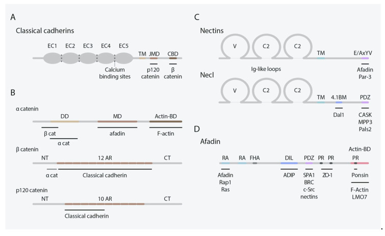
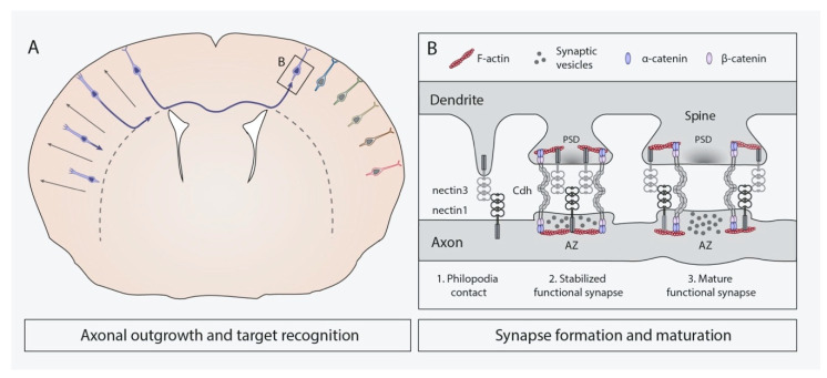
The neocortex is an exquisitely organized structure achieved through complex cellular processes from the generation of neural cells to their integration into cortical circuits after complex migration processes. During this long journey, neural cells need to establish and release adhesive interactions through cell surface receptors known as cell adhesion molecules (CAMs). Several types of CAMs have been described regulating different aspects of neurodevelopment. Whereas some of them mediate interactions with the extracellular matrix, others allow contact with additional cells. In this review, we will focus on the role of two important families of cell-cell adhesion molecules (C-CAMs), classical cadherins and nectins, as well as in their effectors, in the control of fundamental processes related with corticogenesis, with special attention in the cooperative actions among the two families of C-CAMs.
Keywords: CAMs; axon targeting; classical cadherins; nectins; neocortical development; neurodevelopmental disorders; neuronal migration; neurons; radial glia cells; synaptogenesis.
Conflict of interest statement
The authors declare no conflict of interest.
Figures


Similar articles
-
Right Place at the Right Time: How Changes in Protocadherins Affect Synaptic Connections Contributing to the Etiology of Neurodevelopmental Disorders.Cells. 2020 Dec 18;9(12):2711. doi: 10.3390/cells9122711. Cells. 2020. PMID: 33352832 Free PMC article. Review.
-
Nectins and Nectin-like molecules in synapse formation and involvement in neurological diseases.Mol Cell Neurosci. 2021 Sep;115:103653. doi: 10.1016/j.mcn.2021.103653. Epub 2021 Jul 7. Mol Cell Neurosci. 2021. PMID: 34242750 Review.
-
Proteolytic remodeling of the synaptic cell adhesion molecules (CAMs) by metzincins in synaptic plasticity.Neurochem Res. 2013 Jun;38(6):1113-21. doi: 10.1007/s11064-012-0919-6. Epub 2012 Nov 4. Neurochem Res. 2013. PMID: 23124395 Free PMC article. Review.
-
Cell adhesion molecules nectins and associating proteins: Implications for physiology and pathology.Proc Jpn Acad Ser B Phys Biol Sci. 2010;86(6):621-9. doi: 10.2183/pjab.86.621. Proc Jpn Acad Ser B Phys Biol Sci. 2010. PMID: 20551598 Free PMC article. Review.
-
The roles of nectins in cell adhesions: cooperation with other cell adhesion molecules and growth factor receptors.Curr Opin Cell Biol. 2007 Oct;19(5):593-602. doi: 10.1016/j.ceb.2007.09.007. Epub 2007 Oct 17. Curr Opin Cell Biol. 2007. PMID: 17942295 Review.
Cited by
-
Comprehensive analysis of prognostic significance of cadherin (CDH) gene family in breast cancer.Aging (Albany NY). 2022 Oct 30;14(20):8498-8567. doi: 10.18632/aging.204357. Aging (Albany NY). 2022. PMID: 36315446 Free PMC article.
-
Functional characterization of the schizophrenia associated gene AS3MT identifies a role in neuronal development.Am J Med Genet B Neuropsychiatr Genet. 2022 Jul;189(5):151-162. doi: 10.1002/ajmg.b.32905. Epub 2022 Jun 19. Am J Med Genet B Neuropsychiatr Genet. 2022. PMID: 35719055 Free PMC article.
-
Molecular logic for cellular specializations that initiate the auditory parallel processing pathways.Nat Commun. 2025 Jan 9;16(1):489. doi: 10.1038/s41467-024-55257-z. Nat Commun. 2025. PMID: 39788966 Free PMC article.
-
Immunoglobulin superfamily 3 (Igsf3) function is dispensable for brain development.Sci Rep. 2025 Feb 23;15(1):6526. doi: 10.1038/s41598-024-79349-4. Sci Rep. 2025. PMID: 39988603 Free PMC article.
-
A guide to heat shock factors as multifunctional transcriptional regulators.FEBS J. 2025 Aug;292(16):4133-4155. doi: 10.1111/febs.70139. Epub 2025 Jun 2. FEBS J. 2025. PMID: 40457168 Free PMC article. Review.
References
Publication types
MeSH terms
Substances
LinkOut - more resources
Full Text Sources
Other Literature Sources
