. 2021 Jan 10;10(1):120.
doi: 10.3390/cells10010120.

Role of Synovial Exosomes in Osteoclast Differentiation in Inflammatory Arthritis

Affiliations

Role of Synovial Exosomes in Osteoclast Differentiation in Inflammatory Arthritis

Ji Eun Song et al. Cells. .

Abstract

This study aimed to investigate the characteristics of exosomes isolated from synovial fluid and their role in osteoclast differentiation in different types of inflammatory arthritis. Exosomes isolated from synovial fluid of rheumatoid arthritis (RA), ankylosing spondylitis (AS), gout, and osteoarthritis (OA) patients were co-incubated with CD14+ mononuclear cells from healthy donors without macrophage colony-stimulating factor (M-CSF) and receptor activator of nuclear factor kappa-B ligand (RANKL). Osteoclast differentiation was evaluated via tartrate-resistant acid phosphatase (TRAP) staining and activity and F-actin ring formation. RANKL expression on synovial exosomes was assessed using flow cytometry and an enzyme-linked immunosorbent assay (ELISA). Synovial exosomes were the lowest in OA patients; these induced osteoclastogenesis in the absence of M-CSF and RANKL. Osteoclastogenesis was significantly higher with more exosomes in RA (p = 0.030) than in OA patients, but not in AS or gout patients. On treating macrophages with a specified number of synovial exosomes from RA/AS patients, exosomes induced greater osteoclastogenesis in RA than in AS patients. Synovial exosomal RANKL levels were significantly higher in RA (p = 0.035) than in AS patients. Synovial exosome numbers vary with the type of inflammatory arthritis. Synovial exosomes from RA patients may bear the disease-specific "synovial signature of osteoclastogenesis."

Keywords: inflammatory arthritis; osteoclastogenesis; synovial exosomes.


Conflict of interest statement

The authors declare no conflict of interest.

Figures
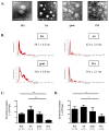
Figure 1
Figure 1
Characterization of synovial exosomes. (A) The shape and size of synovial exosomes purified from rheumatoid arthritis (RA), ankylosing spondylitis (AS), gout, and osteoarthritis (OA) patients were observed using a transmission electron microscope (TEM). The size of synovial exosomes ranged in diameter from 20 to 200 nm. Scale bar = 200 nm. (B) The size distribution of synovial exosomes was measured using a Nanosight LM 10 and analyzed with enzyme-linked immunosorbent assay (NTA) 3.1 software. Red error bars indicate +/−1 standard error of the mean, and the modal size of synovial exosomes are shown for each preparation. (C,D) The number of synovial exosomes was assessed by acetyl-CoA acetylcholinesterase (AChE) activity and CD81-ELISA. Data are presented as the mean ± SEM. * p < 0.05 vs. OA, ** p < 0.01 vs. OA.
Figure 2
Figure 2
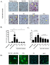
Treatment of synovial exosomes with human macrophages and the effect of synovial exosomes on human osteoclastogenesis. (A) Human macrophages differentiated from CD14+ monocytes were treated with 20 ng/mL of macrophage colony-stimulating factor (M-CSF) + 40 ng/mL of receptor activator of nuclear factor kappa-B ligand (RANKL) (positive control) or with 10% synovial exosomes, respectively. Exosomes were isolated from the same volume of synovial fluid with rheumatoid arthritis (RA), ankylosing spondylitis (AS), gout, and osteoarthritis (OA) patients. After 9–10 days, cells were stained for tartrate-resistant acid phosphatase (TRAP) expression, and TRAP-positive multinucleated cells (MNCs) were imaged using light microscopy. The yellow arrow indicates TRAP-positive MNCs. Magnification: ×100. (B) The number of TRAP-positive multinucleated (more than three) osteoclasts were counted (cells/cm2). Data are presented as a fold change in the osteoclast number compared to medium alone (negative control) and mean ± SEM. ** p < 0.01 vs. medium alone, # p < 0.05 vs. OA. (C) TRAP activity was measured at 405 nm and expressed as fold change in the medium alone. Data are presented as the mean ± SEM. (D) Actin rings were stained with FTIC-phalloidin, and nuclei were stained with Hoechst 33258. The white arrow indicates the F-actin ring. Scale bar = 50 μm.
Figure 3
Figure 3
RANKL expression on synovial exosomes. (A) Exosomes from rheumatoid arthritis (RA) and ankylosing spondylitis (AS) patients were lysed for analysis of RANKL concentration in synovial exosomes. The levels of RANKL were measured using sandwich enzyme-linked immunosorbent assay (ELISA). Data are presented as the mean ± SEM. * p < 0.05 vs. disease control, AS. (B) CD9 or anti-RANKL antibodies-coated magnetic beads and stained with Exo-FITC. The expression of CD9 and RANKL are presented by flow cytometric histograms.

References

    1. Radner H., Ramiro S., Buchbinder R., Landewe R.B., van der Heijde D., Aletaha D. Pain management for inflammatory arthritis (rheumatoid arthritis, psoriatic arthritis, ankylosing spondylitis and other spondylarthritis) and gastrointestinal or liver comorbidity. Cochrane Database Syst. Rev. 2012;1:CD008951. doi: 10.1002/14651858.CD008951.pub2. - DOI - PMC - PubMed
    1. Feldmann M., Brennan F.M., Maini R.N. Role of cytokines in rheumatoid arthritis. Annu. Rev. Immunol. 1996;14:397–440. doi: 10.1146/annurev.immunol.14.1.397. - DOI - PubMed
    1. Miyasaka N., Sato K., Goto M., Sasano M., Natsuyama M., Inoue K., Nishioka K. Augmented interleukin-1 production and HLA-DR expression in the synovium of rheumatoid arthritis patients. Possible involvement in joint destruction. Arthritis Rheum. 1988;31:480–486. doi: 10.1002/art.1780310404. - DOI - PubMed
    1. Goldring S.R., Purdue P.E., Crotti T.N., Shen Z., Flannery M.R., Binder N.B., Ross F.P., McHugh K.P. Bone remodelling in inflammatory arthritis. Ann. Rheum. Dis. 2013;72(Suppl. 2):ii52–ii55. doi: 10.1136/annrheumdis-2012-202199. - DOI - PubMed
    1. Schett G. Joint remodelling in inflammatory disease. Ann. Rheum. Dis. 2007;66(Suppl. 3):iii42–iii44. doi: 10.1136/ard.2007.078972. - DOI - PMC - PubMed

Publication types