Review
. 2021 Jan 9;13(2):217.
doi: 10.3390/cancers13020217.

The Role of Tumor Microenvironment in Multiple Myeloma Development and Progression

Affiliations
Review

The Role of Tumor Microenvironment in Multiple Myeloma Development and Progression

Almudena García-Ortiz et al. Cancers (Basel). .

Abstract

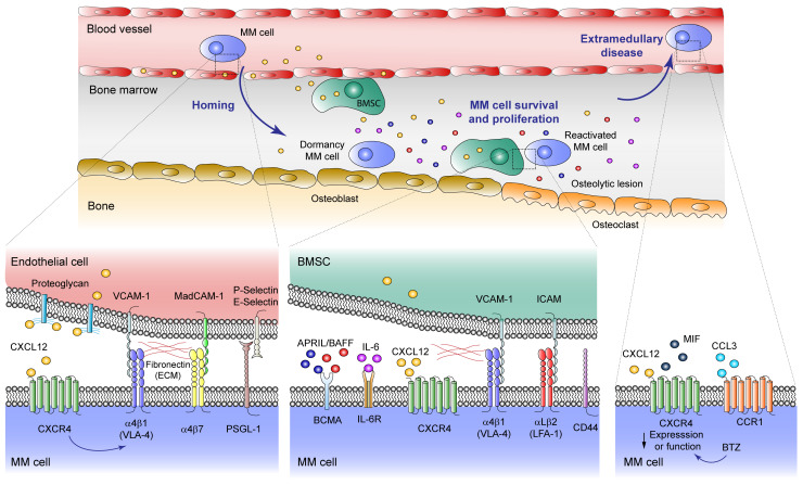
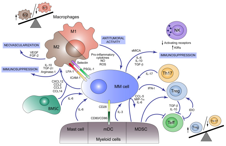
Multiple myeloma (MM) is a hematologic cancer characterized by clonal proliferation of plasma cells in the bone marrow (BM). The progression, from the early stages of the disease as monoclonal gammopathy of undetermined significance (MGUS) and smoldering multiple myeloma (SMM) to MM and occasionally extramedullary disease, is drastically affected by the tumor microenvironment (TME). Soluble factors and direct cell-cell interactions regulate MM plasma cell trafficking and homing to the BM niche. Mesenchymal stromal cells, osteoclasts, osteoblasts, myeloid and lymphoid cells present in the BM create a unique milieu that favors MM plasma cell immune evasion and promotes disease progression. Moreover, TME is implicated in malignant cell protection against anti-tumor therapy. This review describes the main cellular and non-cellular components located in the BM, which condition the immunosuppressive environment and lead the MM establishment and progression.

Keywords: adhesion molecules; bone marrow niche; immunosuppression; migration; multiple myeloma; tumor microenvironment.


Conflict of interest statement

The authors declare no potential conflict of interest.

Figures

Figure 1
Figure 1
Malignant plasma cell trafficking events in multiple myeloma (MM) development. MM plasma cells reach the bone marrow (BM) niche through sinusoids, proliferate favored by the tumor microenvironment (TME) and, occasionally, egress to the circulation causing extramedullary disease. Homing: The interaction of the CXCL12 chemokine with its CXCR4 receptor, mediates homing, lodging and retention of both normal and malignant plasma cells into the BM. CXCL12–CXCR4 interaction upregulates the activity of the α4β1 integrin, allowing high binding to its ligand VCAM-1 expressed on the BM microvasculature. Other important adhesion molecules mediating MM cell homing into the BM are the α4β7 integrin, a receptor for MAdCAM-1 and fibronectin, and PSGL-1 which interacts with P- and E-selectin expressed on the endothelial cell surface. MM cell survival and proliferation: Inside the BM milieu, BM mesenchymal stromal cells (BMSC) secrete high levels of CXCL12 that, along with α4β1, α4β7, and αLβ2 integrins, as well as CD44, are important for anchoring and retention of MM cells into BM niches. During this stage, survival and proliferation of malignant plasma cells is contributed by two main soluble mediators, a proliferation-inducing ligand (APRIL) and B-cell activating factor (BAFF), which bind B-cell maturation antigen (BCMA) in the cancer cell surface, and IL-6, whose receptor is also expressed on MM cells. In MM patients there is a pathological osteolineage imbalance with reduced osteoblasts in favor of osteoclasts which produce lytic lesions. BM osteoblastic niche facilitates the dormancy of MM cells, while osteoclasts induce MM cell reactivation. Extramedullary disease: In this context, cancer cells become independent from the TME, and CXCR4 function or expression is downregulated, an event that also occurs after bortezomib (BTZ) treatment. Macrophage migration inhibitory factor (MIF) can also bind to CXCR4, inducing the expression of adhesion molecules. Expression of CCR1 chemokine receptor is linked to MM plasma cell circulation.
Figure 2
Figure 2
Immunosuppressive tumor microenvironment (TME) in multiple myeloma (MM). Both myeloid and lymphoid cells present in bone marrow (BM) participate in MM development. M1 macrophages act as antitumoral agents producing pro-inflammatory cytokines, nitric oxide (NO) and reactive oxygen species (ROS) and acting as antigen-presenting cells (APC) that activate immune response against MM cells. MM cells and BM mesenchymal stromal cells (BMSCs) produce the chemokines CXCL12, CCL2, CCL3, and CCL14, which promote macrophage M2 polarization and migration to the tumor niche, unbalancing M1/M2 ratio towards the M2 population. M2 macrophages play a pro-tumoral role secreting immunosuppressive agents such as IL-10, TGF-β1 and Arginase-1 and neovascularization factors like VEGF and FGF-2. M2 macrophages act as MM cell protectors through the expression of selectins and LFA-1 in the macrophage surface, which binds to PSGL-1 and ICAM-1 in the plasma cell, respectively. Mast cells, although contributing to an initial tumoricidal host response, secrete IL-6 which promotes MM cell growth. Myeloid dendritic cells (mDCs) foster MM cell proliferation and survival by cell–cell interactions with tumor cells via CD80/CD86-CD28 and secretion of IL-3. Additionally, myeloid-derived suppressor cells (MDSCs) induce the secretion of cytokines such as CCL5, MIP-1α and IL-6 by MM cells and modulate the cytotoxic T cell responses inhibiting effector T cell functions and activating regulatory T cells (Treg). Other signals that promote Treg cell expansion are IFN-I, produced by MM cells, and indoleamine 2, 3-dioxygenase (IDO) activity which at the same time inhibits effector T cells. Cytokines produced by Tregs, such as IL-10 and TGF-β, together with other MM cell-derived signals attenuate effector T cell function. Th17 lymphocytes are also present in BM from MM patients and foster tumor growth through IL-17 secretion. NK cell cytotoxic activity is modulated during disease progression by cytokines and soluble factors (e.g., sMICA) secreted by MM cells that induce a reduction in activating receptors and an increase in inhibitory receptors (KIRs).

Similar articles

Cited by

References

    1. Siegel R.L., Miller K.D., Jemal A. Cancer statistics, 2019. CA A Cancer J. Clin. 2019;69:7–34. doi: 10.3322/caac.21551. - DOI - PubMed
    1. Kumar S.K., Rajkumar V., Kyle R.A., van Duin M., Sonneveld P., Mateos M.V., Gay F., Anderson K.C. Multiple myeloma. Nat. Rev. Dis. Primers. 2017;3:17046. doi: 10.1038/nrdp.2017.46. - DOI - PubMed
    1. Redondo-Munoz J., Garcia-Pardo A., Teixido J. Molecular Players in Hematologic Tumor Cell Trafficking. Front Immunol. 2019;10:156. doi: 10.3389/fimmu.2019.00156. - DOI - PMC - PubMed
    1. Ghobrial I.M. Myeloma as a model for the process of metastasis: Implications for therapy. Blood. 2012;120:20–30. doi: 10.1182/blood-2012-01-379024. - DOI - PMC - PubMed
    1. Chu V.T., Berek C. The establishment of the plasma cell survival niche in the bone marrow. Immunol. Rev. 2013;251:177–188. doi: 10.1111/imr.12011. - DOI - PubMed

LinkOut - more resources