. 2021 Jan 9;22(2):598.
doi: 10.3390/ijms22020598.

K2 Transfection System Boosts the Adenoviral Transduction of Murine Mesenchymal Stromal Cells

Affiliations

K2 Transfection System Boosts the Adenoviral Transduction of Murine Mesenchymal Stromal Cells

Madalina Dumitrescu et al. Int J Mol Sci. .

Abstract

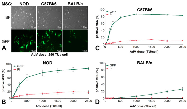
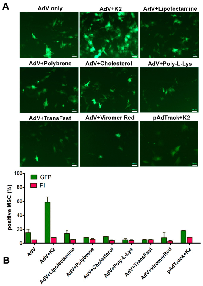
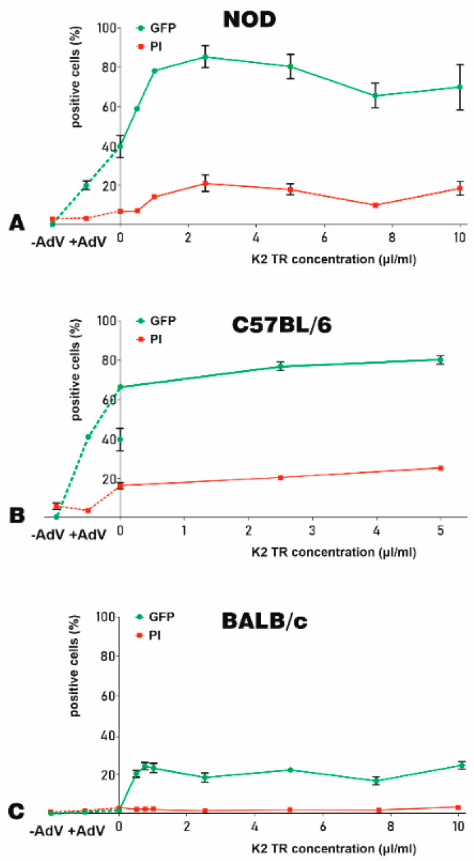
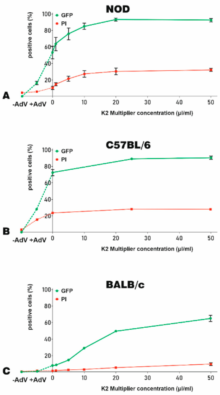
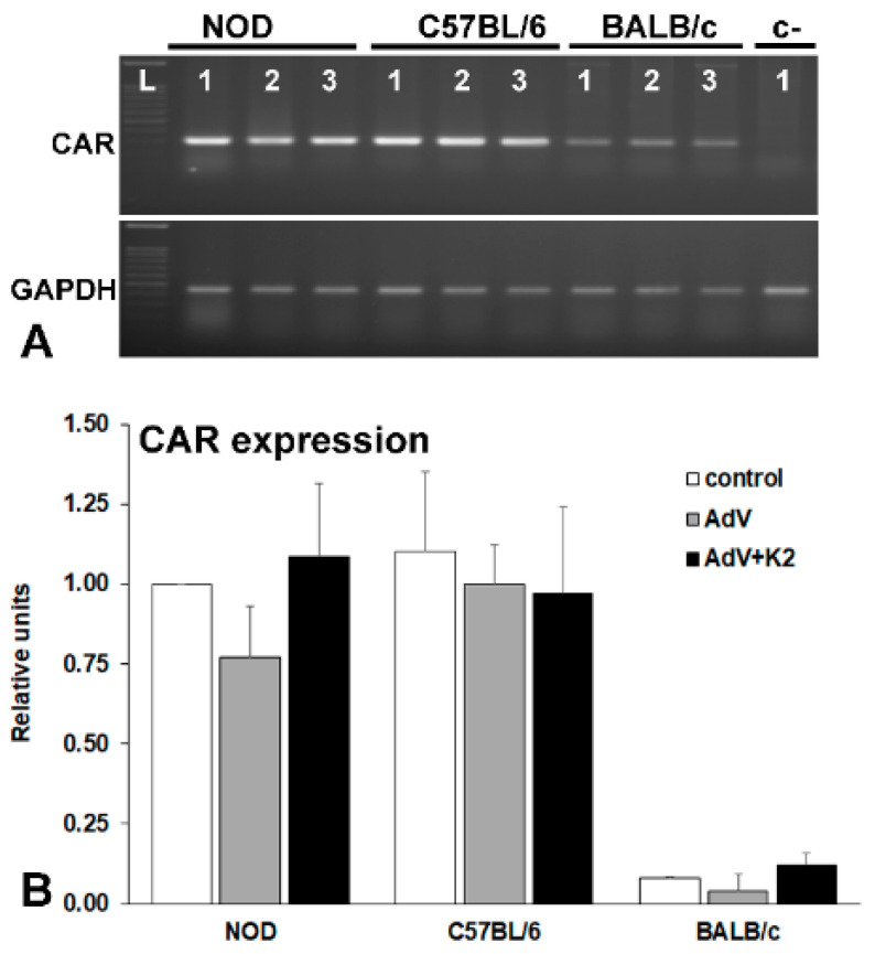
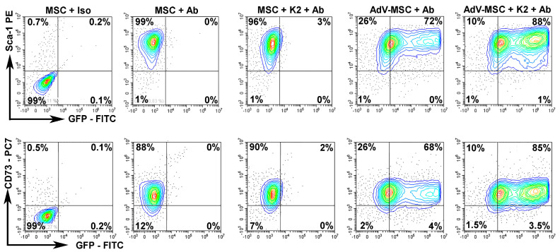
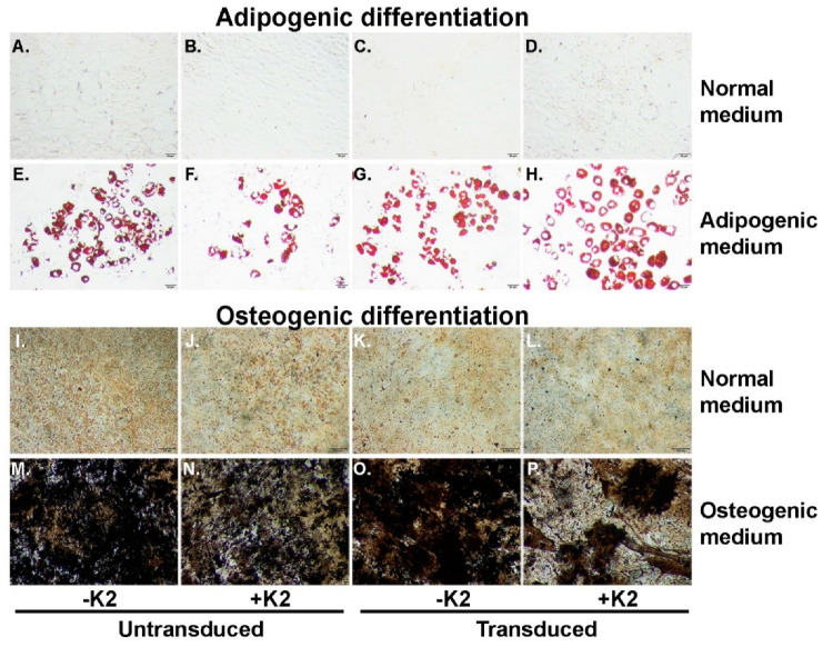
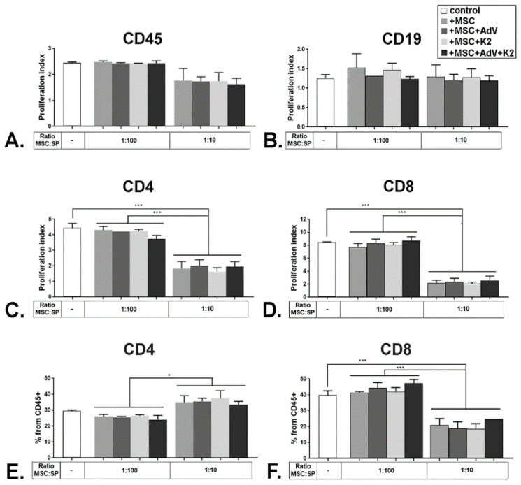
Adenoviral vectors are important vehicles for delivering therapeutic genes into mammalian cells. However, the yield of the adenoviral transduction of murine mesenchymal stromal cells (MSC) is low. Here, we aimed to improve the adenoviral transduction efficiency of bone marrow-derived MSC. Our data showed that among all the potential transduction boosters that we tested, the K2 Transfection System (K2TS) greatly increased the transduction efficiency. After optimization of both K2TS components, the yield of the adenoviral transduction increased from 18% to 96% for non-obese diabetic (NOD)-derived MSC, from 30% to 86% for C57BL/6-derived MSC, and from 0.6% to 63% for BALB/c-derived MSC, when 250 transduction units/cell were used. We found that MSC derived from these mouse strains expressed different levels of the coxsackievirus and adenovirus receptors (MSC from C57BL/6≥NOD>>>BALB/c). K2TS did not increase the level of the receptor expression, but desensitized the cells to foreign DNA and facilitated the virus entry into the cell. The expression of Stem cells antigen-1 (Sca-1) and 5'-nucleotidase (CD73) MSC markers, the adipogenic and osteogenic differentiation potential, and the immunosuppressive capacity were preserved after the adenoviral transduction of MSC in the presence of the K2TS. In conclusion, K2TS significantly enhanced the adenoviral transduction of MSC, without interfering with their main characteristics and properties.

Keywords: BALC/c-derived MSC; C57BL/6-derived MSC; K2 transfection system; NOD-derived MSC; adenovirus; mesenchymal stromal cells; transduction.


Conflict of interest statement

The authors declare no conflict of interest. The funders had no role in the design of the study; in the collection, analyses, or interpretation of data; in the writing of the manuscript, or in the decision to publish the results.

Figures

Figure 1
Figure 1
Adenoviral transduction efficiency of murine mesenchymal stromal cells (MSC) isolated from three mouse strains. (A) Microscopy of non-obese diabetic NOD-MSC, C57BL/6-MSC, and BALB/c-MSC, transduced with 250 transduction units (TU)/cell adenovirus for GFP expression: BF—Bright field and GFP—fluorescence microscopy. (BD) Dose-dependent transduction of MSC derived from NOD, C57BL/6, and BALB/c strains. The cells were incubated with increasing doses of adenovirus ranging from 0–2500 TU/cell; after 48 h the GFP expressing cells and the cell death determined by propidium iodide (PI) incorporation were evaluated by flow cytometry. The dose-dependent curves are different for NOD-MSC (B), C57BL/6-MSC (C), and BALB/c-MSC (D).
Figure 2
Figure 2
Adenoviral transduction of murine mesenchymal stromal cells in the presence of different potential transduction boosters. To induce GFP expression, MSC were incubated with 250 TU/cell adenovirus alone (AdV only) or in the presence of the K2 Transfection System (K2TS) (AdV + K2), Lipofectamine 3000 (AdV + Lipofectamine), 10 μg/mL Polybrene (AdV + Polybrene), 2 μg/mL free cholesterol (AdV + Cholesterol), 1 μg/mL poly-L-Lysine (AdV + Poly-L-Lys), TransFast (AdV + TransFast), or Viromer Red (AdV + ViromerRed). In parallel, MSC were transfected with pAdTrack-CMV using K2TS (pAdTrack + K2). After 48 h, the GFP expression was observed by fluorescence microscopy (A), and the number of the GFP-expressing cells was determined by flow cytometry (B). The percentage of GFP-positive cells determined by flow cytometry is represented by green columns and that of the dead cells colored with propidium iodide (PI)—in red columns. As is revealed, the K2TS is the most efficient reagent for boosting the adenoviral transduction of MSC. Bars, 20 µm.
Figure 3
Figure 3
K2 Transfection Reagent K2TR optimization for the adenoviral transduction of murine MSC. The efficacy of K2TR to increase the yield of the adenoviral transduction of MSC derived from NOD (A), C57BL/6 (B), and BALB/c (C) mice were determined as % of GFP-positive cells (green lines). The cell death was determined by PI incorporation and expressed as % of PI-positive cells (red lines). On the left side of each graph, the GFP-positive cells and the cell death for the untransduced cells (−AdV) and for the cells transduced with 250 TU/cell adenovirus alone (+AdV) were illustrated, linked by a dotted line. Adenoviral particles were complexed with various K2TR doses. MSC were incubated with 10 μL/mL K2 Multiplier (K2M) and then the complexes of adenovirus–K2TR were added to the cells. The percentage of GFP-positive cells (green lines) induced by transduction and the percentage of PI-positive cells (red lines) were determined 48 h after transduction, by flow cytometry.
Figure 4
Figure 4
The optimization of K2M for adenoviral transduction of murine MSC. The efficacy of K2M to increase the adenoviral transduction of MSC-derived from NOD (A), C57BL/6 (B), and BALB/c (C) mice were expressed as % of GFP-positive cells (green lines). The K2M induced cytotoxicity was indicated by the % of PI-positive, dead cells (red lines). To optimize the concentration of K2M needed for transduction, MSC were incubated with increasing K2M concentrations (up to 50 μL/mL) for 90 min. Then, the adenovirus–K2TR complexes (5 μL/mL K2TR and 250 TU/cell adenoviral particle) were added to the cells. The percentage of GFP-positive cells and PI-positive cells at 48 h after transduction was determined by flow cytometry. The same fractions were evidenced also for untransduced cells (−AdV) and NOD-, C57BL/6- and BALB/c-MSC transduced with the adenovirus alone (+AdV), which were linked by a dotted line in the left side of the graphs.
Figure 5
Figure 5
Evaluation of coxsackievirus and adenovirus receptor (CAR) expression in MSC derived from NOD, C57BL/6, and BALB/c mice analyzed by RT-PCR (A) and by Q-PCR (B). CAR expression is higher in NOD- and C57BL/6-MSC, as compared with that in BALB/c-MSC (A, lane 1 and B, white columns) and is not modified by the adenovirus (A, lane 2 and B, grey columns) or by the K2TS (A, lane 3 and B. black columns).
Figure 6
Figure 6
K2TS does not alter the markers expressed by MSC. The expression of Stem cells antigen-1 (Sca-1) and 5′-nucleotidase (CD73) was tested in NOD-MSC by flow cytometry. MSC were incubated with the corresponding isotype antibody (MSC + Iso) to set the gates. MSC were incubated with the corresponding antibodies (MSC + Ab) or were treated with K2TS and incubated with the antibodies (MSC + K2 + Ab). In parallel, MSC were transduced with the adenovirus alone (AdV-MSC + Ab) or in the presence of K2TS (AdV-MSC + K2 + Ab). In the upper panel the specific antibodies were anti-Sca-1 and in the lower panel were anti-CD73 antibodies. Both transduced and untransduced cells expressed a high amount of the markers (~98%). The GFP expression was detected in 16–20% of the cells transduced with the adenovirus alone (AdV-MSC + Ab) and in 90 - 92% in cells transduced with the adenovirus together with the K2TS (AdV-MSC + K2 + Ab).
Figure 7
Figure 7
Exposure to K2TS does not influence the multipotency of MSC. Unmanipulated cells (A,E,I,M), K2TS-treated, untransduced (B,F,J,N), AdV-transduced (C,G,K,O), and AdV-transduced in the presence of K2TS (D,H,L,P) were cultured in normal MSC medium (AD) and (IL), or in adipogenic medium (EH) or in osteogenic medium (MP) for two weeks. The cells treated as described above were stained with Oil Red O (AH), or with von Kossa protocol (IP). Bars are 50 µm (AH) and 200 µm (IP).
Figure 8
Figure 8
Immunomodulatory properties of MSC are not affected upon exposure to KTS. Whole splenocytes were labeled with carboxy-fluorescein succinimidyl ester (CFSE) and cultured in the presence of CD3/CD28 stimulation beads for 72 h alone (control), or together with naïve MSC (+MSC), transduced MSC (+MSC + AdV), K2TS-treated MSC (+MSC + K2) and transduced MSC in the presence of K2TS (+MSC + AdV + K2), in different ratios, as indicated. At the end of the incubation time, the proliferation index of the populations of splenocytes positive for CD45 (A), CD19 (B), CD4 (C), and CD8 (D) was calculated. The percentage of CD4+ (E) and CD8+ (F) cells from the CD45 positive splenocytes were determined and represented. * corresponds to a p-value < 0.05, and *** for p < 0.0005, by ANOVA.

Similar articles

Cited by

References

    1. Haynesworth S.E., Goshima J., Goldberg V.M., Caplan A.I. Characterization of cells with osteogenic potential from human marrow. Bone. 1992;13:81–88. doi: 10.1016/8756-3282(92)90364-3. - DOI - PubMed
    1. Pittenger M.F., Mackay A.M., Beck S.C., Jaiswal R.K., Douglas R., Mosca J.D., Moorman M.A., Simonetti D.W., Craig S., Marshak D.R. Multilineage potential of adult human mesenchymal stem cells. Science. 1999;284:143–147. doi: 10.1126/science.284.5411.143. - DOI - PubMed
    1. Halvorsen Y.C., Wilkison W.O., Gimble J.M. Adipose-derived stromal cells--their utility and potential in bone formation. Int. J. Obes. Relat. Metab. Disord. 2000;24(Suppl. 4):S41–S44. doi: 10.1038/sj.ijo.0801503. - DOI - PubMed
    1. Zuk P.A., Zhu M., Mizuno H., Huang J., Futrell J.W., Katz A.J., Benhaim P., Lorenz H.P., Hedrick M.H. Multilineage cells from human adipose tissue: Implications for cell-based therapies. Tissue Eng. 2001;7:211–228. doi: 10.1089/107632701300062859. - DOI - PubMed
    1. In ‘t Anker P.S., Scherjon S.A., Kleijburg-van der Keur C., De Groot-Swings G.M., Claas F.H., Fibbe W.E., Kanhai H.H. Isolation of mesenchymal stem cells of fetal or maternal origin from human placenta. Stem Cells. 2004;22:1338–1345. doi: 10.1634/stemcells.2004-0058. - DOI - PubMed

Substances