. 2021 Jan 9;7(1):38.
doi: 10.3390/jof7010038.

Genetic Manipulation of the Brassicaceae Smut Fungus Thecaphora thlaspeos

Affiliations

Genetic Manipulation of the Brassicaceae Smut Fungus Thecaphora thlaspeos

Lesley Plücker et al. J Fungi (Basel). .

Abstract

Investigation of plant-microbe interactions greatly benefit from genetically tractable partners to address, molecularly, the virulence and defense mechanisms. The smut fungus Ustilago maydis is a model pathogen in that sense: efficient homologous recombination and a small genome allow targeted modification. On the host side, maize is limiting with regard to rapid genetic alterations. By contrast, the model plant Arabidopsis thaliana is an excellent model with a vast amount of information and techniques as well as genetic resources. Here, we present a transformation protocol for the Brassicaceae smut fungus Thecaphora thlaspeos. Using the well-established methodology of protoplast transformation, we generated the first reporter strains expressing fluorescent proteins to follow mating. As a proof-of-principle for homologous recombination, we deleted the pheromone receptor pra1. As expected, this mutant cannot mate. Further analysis will contribute to our understanding of the role of mating for infection biology in this novel model fungus. From now on, the genetic manipulation of T. thlaspeos, which is able to colonize the model plant A. thaliana, provides us with a pathosystem in which both partners are genetically amenable to study smut infection biology.

Keywords: homologous recombination; infection; mating; pheromone receptor; protoplast; smut; transformation.


Conflict of interest statement

The authors declare no conflict of interest. The funders had no role in the design of the study; in the collection, analyses, or interpretation of data; in the writing of the manuscript, or in the decision to publish the results.

Figures

Figure 1
Figure 1
Identification of an osmotic stabilizer. Thecaphora thlaspeos LF1 culture was grown to an OD600 = 0.4–0.8. To optimize protoplasting of T. thlaspeos hyphae by Yatalase and Glucanex, the osmotic stabilizers MgSO4, sorbitol, and sucrose were tested. With MgSO4 as osmotic stabilizer, all filaments were digested; while in sorbitol and sucrose, no protoplasts were obtained. Black arrowheads: filaments; white arrowheads: protoplast; scale bar: 50 µm.
Figure 2
Figure 2
Protoplasting filamentous T. thlaspeos cultures. 1–2 g fresh weight of an exponentially growing culture (A) was harvested by filtration and protoplasted with Glucanex and Yatalase for 30–60 min. To purify the crude protoplasts (B), they are floated in a gradient (C). Filaments and debris are found in the pellet, the protoplasts can be collected from the interphase (marked with a white box). (D) An efficient protoplasting reaction results in up to 108 protoplasts/g fresh weight. Scale bar: 10 µm.
Figure 3
Figure 3
Optimizing the enzyme cocktail for protoplasting. (A) Filamentous cultures were harvested and incubated with a combination of 20 mg/mL Glucanex and 10 mg/mL Yatalase, or each enzyme individually, for 30 min. Protoplasting was efficient only when both enzymes were applied. (B) The enzymes were diluted to identify the lowest suitable concentration. The frequency of remaining filaments is inversely proportional to the enzyme concentration. The highest efficiency was obtained with 20 mg/mL Glucanex and 10 mg/mL Yatalase. White arrowhead: protoplasts; black arrowhead: residual filaments; scale bar: 10 µm.
Figure 4
Figure 4
Optimizing protoplast regeneration. (A) Protoplasts regenerated for 39 days on regeneration medium (YMPG) with either 1 M sorbitol, sucrose, glucose, or KCl. No regeneration was observed without osmotic stabilizer. It was most efficient on sucrose, followed by glucose and sorbitol, while KCl inhibited cell growth. (B) T. thlaspeos LF1 culture plated on YMPG with sorbitol, sucrose, glucose, KCl, or without osmotic stabilizer. After 21 days, no growth was observed for 1 M KCl. Growth rate is reduced for 1 M sorbitol, glucose, and sucrose compared to absence of osmotic stabilizer. (C) Regeneration of protoplasts on YMPG with 1 M sucrose documented microscopically for 11–18 days. Initially, the protoplasts turned dark (2 days) before new filaments emerged (3–8 days). The filaments proliferated (11–18 days), resulting in macroscopically visible colonies (28–35 days). White arrowhead: protoplasts; black arrowhead: emerging filaments; scale bar: 10 µm.
Figure 5
Figure 5
Verification of resistance-reporter constructs in U. maydis. Reporter constructs containing a fusion of hygromycin-phospho-transferase gene (hpt) and the fluorescent marker (egfp or mcherry) under the control of hsp70 promoter and terminator regions derived from the T. thlaspeos genome were tested in U. maydis. Upon transformation of the linearized construct, it randomly integrates into the genome. Protein accumulation was visualized by the green/red fluorescence. The eGfp expression under the promoter region of T. thlaspeos was stronger than compared to the stably integrated construct under the control of a strong, synthetic promoter (Potef). This confirms that the fusion protein is active. In comparison, mcherry-fluorescence in the strain carrying the Tthsp70 promoter was weaker than the stably integrated construct under the control of the Potef promoter. Scale bar: 10 µm.
Figure 6
Figure 6
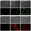
Generation of reporter lines in T. thlaspeos. Reporter constructs containing the active fusion of the hygromycin-phospho-transferase gene (hpt) and the fluorescent marker (egfp or mcherry) under the control of the strong hsp70 promoter from T. thlaspeos were transformed into the cultures T. thlaspeos LF1 or LF2, respectively. Fluorescent signals accumulate in the cytosol of all cells. Strains: hpt-egfp: LF1_PTthsp70::hpt-egfp, and hpt-mcherry: LF2_PTthsp70::hpt-mcherry. Scale bar: 10 µm.
Figure 7
Figure 7
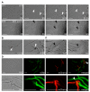
Mating in T. thlaspeos. (A) Top: Confrontation assay of mating partners mfa1/pra1 (LF1) or mfa2/pra2 (LF2). Over the course of 13.5 hours, several mating events of LF1 and LF2 were observed. White arrowheads mark the fusion event. Bottom: Confrontation assay of LF1 pra1∆ and LF2. Over the course of 14 h, hyphae did not mate. Two spots where hyphae could no longer sense each other and cross are marked with black arrow heads. Scale bar: 25 µm. (B,C) Magnification of the mating and crossing events from the white boxes marked in (A). Scale bar: 100 µm. (D) Liquid mating assay with mating partners expressing either cytoplasmic eGfp (LF1-eGfp) or mCherry (LF2-mCherry). Fused hyphae express both eGfp and mCherry and appear yellow in the merged picture (white arrowhead). Scale bar: 25 µm.

References

    1. Kronstad J.W. Pathogenesis and sexual development of the smut fungi. In: Stacey G.K.N.T., editor. Plant-Microbe Interactions. Volume 1 Springer; Boston, MA, USA: 1997.
    1. Feldbrügge M., Kämper J., Steinberg G., Kahmann R. Regulation of mating and pathogenic development in Ustilago maydis. Curr. Opin. Microbiol. 2004;7:666–672. doi: 10.1016/j.mib.2004.10.006. - DOI - PubMed
    1. Brachmann A., König J., Julius C., Feldbrügge M. A reverse genetic approach for generating gene replacement mutants in Ustilago maydis. Mol. Genet. Genom. 2004;272:216–226. doi: 10.1007/s00438-004-1047-z. - DOI - PubMed
    1. Banuett F. Ustilago maydis, the delightful blight. Trends Genet. 1992;8:174–180. doi: 10.1016/0168-9525(92)90220-X. - DOI - PubMed
    1. Vollmeister E., Schipper K., Baumann S., Haag C., Pohlmann T., Stock J., Feldbrügge M. Fungal development of the plant pathogen Ustilago maydis. Fems. Microbiol. Rev. 2012;36:59–77. doi: 10.1111/j.1574-6976.2011.00296.x. - DOI - PubMed

LinkOut - more resources