

Functional T Cell Reactivity to Melanocyte Antigens Is Lost during the Progression of Malignant Melanoma, but Is Restored by Immunization
- PMID: 33435427
- PMCID: PMC7827050
- DOI: 10.3390/cancers13020223
Functional T Cell Reactivity to Melanocyte Antigens Is Lost during the Progression of Malignant Melanoma, but Is Restored by Immunization
Abstract
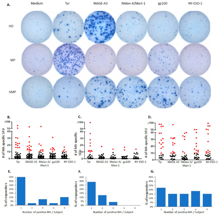
Healthy human subjects develop spontaneous CD8+ T cell responses to melanoma associated antigens (MA) expressed by normal melanocytes, such as Tyrosinase, MAGE-A3, Melan/Mart-1, gp100, and NY-ESO-1. This natural autoimmunity directed against melanocytes might confer protection against the development of malignant melanoma (MM), where MA are present as overexpressed tumor-associated antigens. Consistent with this notion we report here that functional T cell reactivity to MA was found to be significantly diminished to MAGE-A3, Melan-A/Mart-1, and gp100 in untreated MM patients. Three lines of evidence suggest that the MA-reactive T cells present in healthy subjects undergo exhaustion once MM establishes itself. First, only the MA-specific T cell reactivity was affected in the MM patients; that to third party recall antigens was not. Second, in these patients, the residual MA-specific T cells, unlike third party antigen reactive T cells, were functionally impaired, showing a diminished per cell IFN-γ productivity. Third, we show that immunization with MA restored natural CD8+ T cell autoimmunity to MA in 85% of the MM patients. The role of natural T cell autoimmunity to tumor-associated MA is discussed based on discrete levels of T cell activation thresholds.
Keywords: CD8+ T cells; ELISPOT; genetic whole-cell therapeutic melanoma vaccine (AGI-101H); melanoma; melanoma antigens.
Conflict of interest statement
P.V.L., T.Z., A.A.L., and G.A.K. are employees of Cellular Technology Limited (CTL), a company that specializes in ELISPOT testing, producing high-throughput-suitable readers, test kits, and GLP-compliant contract research. A.M., A.P., J.M. and Ł.G., are employees of Poznan University of Medical Sciences. A.M. and Ł.G. are employees of Greater Poland Cancer Centre, Poznan, Poland as well. The Polish authors declare no financial, commercial or other relationships that might be perceived by the academic community as representing a potential conflict of interest.
Figures
References
LinkOut - more resources
Full Text Sources
Other Literature Sources
Research Materials
