. 2021 Jan 9;10(2):219.
doi: 10.3390/jcm10020219.

Animal Model Dependent Response to Pentagalloyl Glucose in Murine Abdominal Aortic Injury

Affiliations

Animal Model Dependent Response to Pentagalloyl Glucose in Murine Abdominal Aortic Injury

Jennifer L Anderson et al. J Clin Med. .

Abstract

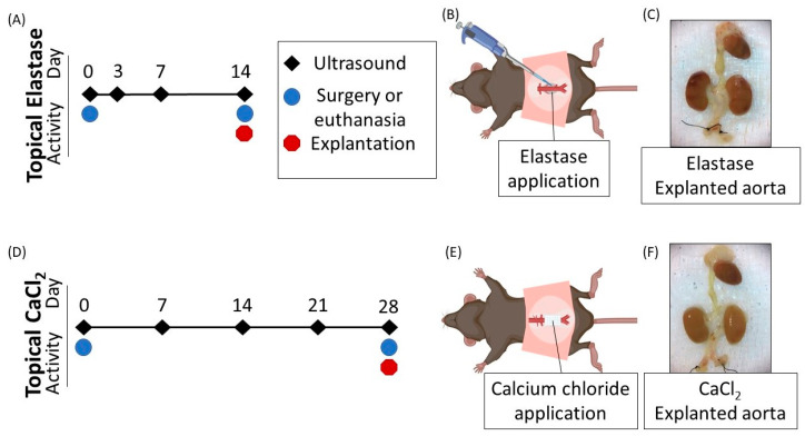
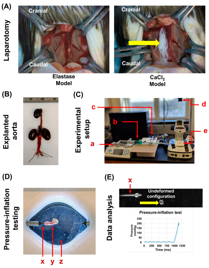
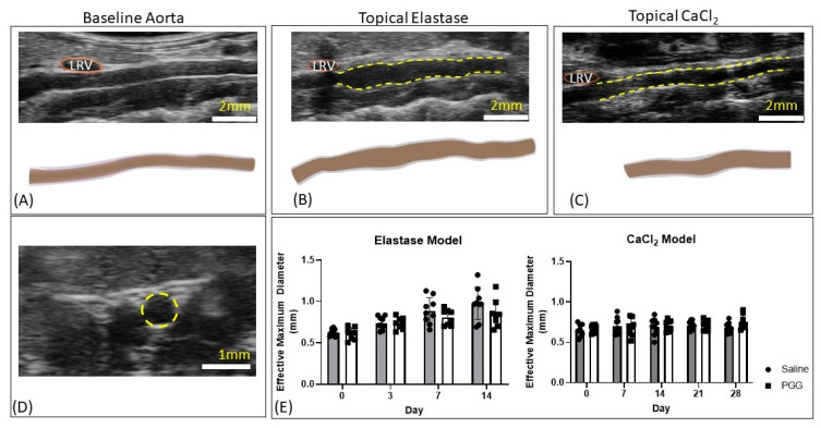
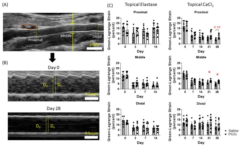
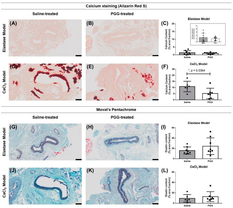
Abdominal aortic aneurysms (AAAs) are a local dilation of the aorta and are associated with significant mortality due to rupture and treatment complications. There is a need for less invasive treatments to prevent aneurysm growth and rupture. In this study, we used two experimental murine models to evaluate the potential of pentagalloyl glucose (PGG), which is a polyphenolic tannin that binds to and crosslinks elastin and collagen, to preserve aortic compliance. Animals underwent surgical aortic injury and received 0.3% PGG or saline treatment on the adventitial surface of the infrarenal aorta. Seventeen mice underwent topical elastase injury, and 14 mice underwent topical calcium chloride injury. We collected high-frequency ultrasound images before surgery and at 3-4 timepoints after. There was no difference in the in vivo effective maximum diameter due to PGG treatment for either model. However, the CaCl2 model had significantly higher Green-Lagrange circumferential cyclic strain in PGG-treated animals (p < 0.05). While ex vivo pressure-inflation testing showed no difference between groups in either model, histology revealed reduced calcium deposits in the PGG treatment group with the CaCl2 model. These findings highlight the continued need for improved understanding of PGG's effects on the extracellular matrix and suggest that PGG may reduce arterial calcium accumulation.

Keywords: abdominal aortic aneurysms; elastin; pentagalloyl glucose; ultrasound.


Conflict of interest statement

C.J.G. is a paid consultant of FUJIFILM VisualSonics. The funders had no role in the design of the study; in the collection, analyses, or interpretation of data; in the writing of the manuscript, or in the decision to publish the results.

Figures

Figure 1
Figure 1
Timeline of experimental events. (A) Timeline for injuries induced by topical elastase. (B) Porcine pancreatic elastase was topically applied by pipette to the infrarenal aorta. (C) A representative aorta from the topical elastase group. Aortas were mechanically tested following explantation. (D) Timeline for injuries induced by calcium chloride (CaCl2). (E) CaCl2 was applied via gauze soaked in 200 µL of 0.5 M CaCl2. (F) A representative aorta from the topical CaCl2 group. Aortas were mechanically tested following explantation.
Figure 2
Figure 2
Biomechanical testing of abdominal aorta specimens. (A) Frontal views of the abdominal cavity of the elastase and CaCl2 models after laparotomy, respectively. The yellow arrow on the CaCl2 model indicates the CaCl2-soaked gauze that was applied to the periadventitial surface of the exposed abdominal aorta to initiate aortic injury. (B) Exemplary image of an explanted intact aorta along with the iliac arteries securely tied with sutures. (C) Experimental setup for pressure-inflation testing. The components are as follows: (a) Chemyx syringe pump, (b) syringe filled with PBS + food dye (for leak detection), (c) Omega pressure transducer with a range of 5 psi, (d) Basler high-resolution monochromatic camera for capturing specimen images, and (e) organ bath with 0.5 mm diameter cannula (World Precision Instruments). (D) Top view (as seen from the Basler camera) of the infrarenal aortic specimen (y), loaded onto the cannula (x), and placed in the saline filled organ bath (z). (E) To measure the thickness of the specimens, aortic rings were dissected (yellow arrow) and placed upright in the organ bath; the cannula (x) was utilized as a reference for image resolution, and the undeformed configuration (transverse view) was captured using the same camera setup. Using a custom LabVIEW script, incremental pressure changes with their accompanied time points were recorded for each specimen.
Figure 3
Figure 3
Diameter changes in the topical elastase and topical CaCl2 models. (A) Longitudinal ultrasound image and three-dimensional rendering of the baseline infrarenal aorta. LRV, left renal vein. (B) Representative longitudinal ultrasound image and three-dimensional rendering of an aorta from the topical elastase group 14 days after aortic injury. Yellow dashed lines indicate where treatment, either saline or PGG, was applied. (C) Representative longitudinal ultrasound image and three-dimensional rendering of an aorta from the topical CaCl2 group 28 days after aortic injury. Yellow dashed lines indicate where treatment, either saline or PGG, was applied. (D) Example short axis ultrasound image used to measure luminal area in systole and diastole to assess effective maximum diameter, as shown in Equation (1). (E) No differences between saline-treated and PGG-treated vessels were seen in either the elastase or CaCl2-treated vessels. Elastase-treated vessels increased in diameter over the two-week observation period.
Figure 4
Figure 4
Green–Lagrange circumferential cyclic strain analysis. (A) M-mode acquisitions were taken at the proximal, middle, and distal infrarenal aorta. Representative locations are shown. LRV, left renal vein. (B) M-mode acquisitions at day 0 and at day 28 in the CaCl2-injured model demonstrating reduced compliance and measurement of the diastolic and systolic luminal diameters used to calculate Green–Lagrange circumferential cyclic strain, Equation (2). Systolic and diastolic measurements were made in triplicate and averaged, and the averages used to calculate Green–Lagrange circumferential cyclic strain. Dd, diastolic diameter. Ds, systolic diameter. (C) Green–Lagrange circumferential cyclic strain at each location in the topical elastase and CaCl2 groups. No difference in strain between saline or PGG treatment was seen in the topical elastase group. A statistically significant difference in strain was seen between treatments at the middle location in the CaCl2 group. Strain between treatments approached significance at the proximal location in the topical CaCl2 group.
Figure 5
Figure 5
Ex vivo mechanical characterization of murine abdominal aortas. Biomechanical effect of PGG on elastase (A) and CaCl2 (B) injury models. No significant differences (p > 0.05 for both cases) were observed between the burst pressure (mmHg) vs. time-to-failure (s) profiles of control and PGG-treated specimens for both elastase and CaCl2 cohorts, respectively.
Figure 6
Figure 6
Histological characterization of abdominal aorta specimens obtained from elastase and CaCl2-treated animal models of aortic injury. Alizarin Red S-based staining and semi-quantitative estimation of calcium (dark red) in specimens from the elastase (AC; inset is the zoomed in view of Figure 6C) and CaCl2 model (DF). Movat’s Pentachrome stained sections and their respective semi-quantifications for elastin (dark violet to black) from all four groups, elastase + saline vs. elastase + PGG (GI) and CaCl2 + saline vs. CaCl2 + PGG (JL), are shown here. Calcium content was found to be significantly lower (almost twice) in the PGG-treated cohort as compared to their saline counterpart in the CaCl2-injured group (p = 0.0364), with almost no change in the elastase groups (p > 0.05; (C,F)). Elastin content was not significantly different between the saline-treated vs. PGG-treated cohorts of either injury models (p > 0.05; (I,L)). Scale bar = 100 µm.

Similar articles

Cited by

References

    1. Underlying Cause of Death, 1999–2015 Results Form. [(accessed on 27 August 2017)]; Available online: https://wonder.cdc.gov/controller/datarequest/D76;jsessionid=ED3A4ABA7F9....
    1. Benjamin E.J.M., Blaha M.J.M., Chiuve S.E.S., Cushman M.M., Das S.R.M., Deo R.M., de Ferranti S.D.M., Floyd J.M., Fornage M., Gillespie C.M., et al. Heart Disease and Stroke Statistics-2017 Update: A Report from the American Heart Association. Circulation. 2017;135 doi: 10.1161/CIR.0000000000000485. - DOI - PMC - PubMed
    1. Nicholls S.C., Gardner J.B., Meissner M.H., Johansen K.H. Rupture in small abdominal aortic aneurysms. J. Vasc. Surg. 1998;28:884–888. doi: 10.1016/S0741-5214(98)70065-5. - DOI - PubMed
    1. Wilmink T.B.M., Quick C.R.G., Hubbard C.S., Day N.E. The influence of screening on the incidence of ruptured abdominal aortic aneurysms. J. Vasc. Surg. 1999;30:203–208. doi: 10.1016/S0741-5214(99)70129-1. - DOI - PubMed
    1. Grant S.W., Grayson A.D., Purkayastha D., Wilson S.D., McCollum C. Logistic risk model for mortality following elective abdominal aortic aneurysm repair. BJS Br. J. Surg. 2011;98:652–658. doi: 10.1002/bjs.7463. - DOI - PubMed

LinkOut - more resources