Review
. 2021 Jan 9;22(2):623.
doi: 10.3390/ijms22020623.

Molecular Mechanisms of Glucocorticoid-Induced Insulin Resistance

Affiliations
Review

Molecular Mechanisms of Glucocorticoid-Induced Insulin Resistance

Carine Beaupere et al. Int J Mol Sci. .

Abstract

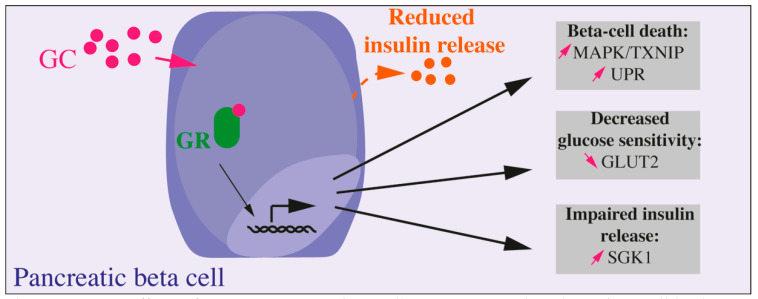
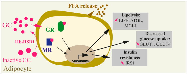
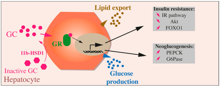
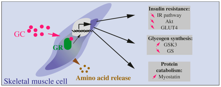
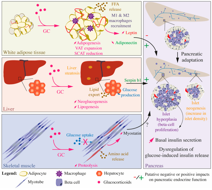
Glucocorticoids (GCs) are steroids secreted by the adrenal cortex under the hypothalamic-pituitary-adrenal axis control, one of the major neuro-endocrine systems of the organism. These hormones are involved in tissue repair, immune stability, and metabolic processes, such as the regulation of carbohydrate, lipid, and protein metabolism. Globally, GCs are presented as 'flight and fight' hormones and, in that purpose, they are catabolic hormones required to mobilize storage to provide energy for the organism. If acute GC secretion allows fast metabolic adaptations to respond to danger, stress, or metabolic imbalance, long-term GC exposure arising from treatment or Cushing's syndrome, progressively leads to insulin resistance and, in fine, cardiometabolic disorders. In this review, we briefly summarize the pharmacological actions of GC and metabolic dysregulations observed in patients exposed to an excess of GCs. Next, we describe in detail the molecular mechanisms underlying GC-induced insulin resistance in adipose tissue, liver, muscle, and to a lesser extent in gut, bone, and brain, mainly identified by numerous studies performed in animal models. Finally, we present the paradoxical effects of GCs on beta cell mass and insulin secretion by the pancreas with a specific focus on the direct and indirect (through insulin-sensitive organs) effects of GCs. Overall, a better knowledge of the specific action of GCs on several organs and their molecular targets may help foster the understanding of GCs' side effects and design new drugs that possess therapeutic benefits without metabolic adverse effects.

Keywords: adipose tissue; glucocorticoids; insulin resistance; liver; muscle; pancreatic beta cells; signaling pathway.


Conflict of interest statement

The authors declare no conflict of interest.

Figures

Figure 1
Figure 1
Direct effects of GC on pancreatic beta cell. GC exposure directly on beta cell leads to altered beta cell function, decreased glucose sensitivity, and insulin secretion and, in fine, to beta cell death. Most of the presented results were obtained in vitro, on isolated islets, primary beta cells, or beta cell lines.
Figure 2
Figure 2
Effects of GC on adipocytes. Inactive GC are activated through the action of the specific enzyme 11b-HSD1. GCs can then bind to their receptors (GC receptor, or GR) or to the mineralocorticoid receptor (MR). The overall action of GCs on mature adipocytes leads to an increase in lipolysis (augmented LIPE, ATGL, and MGLL), a decrease in glucose uptake (decreased GLUT1 and GLUT4) and insulin resistance (reduced IRS1).
Figure 3
Figure 3
Effects of GCs on hepatocytes. Inactive GCs are activated through the action of 11b-HSD1. GCs binding to their receptor (GC receptor, or GR) leads in hepatocytes to glucose production through upregulation of neoglucogenesis (augmented PEPK and G6Pase), to lipids export and insulin resistance through reduced IR pathway, Akt activation, and increased FOXO1.
Figure 4
Figure 4
Effects of GCs on skeletal muscle cells. GCs bind to their receptor (GC receptor, or GR) and lead to protein catabolism (involving increased myostatin production) and amino acids release. Glycogen synthesis is reduced through increased GSK3 and reduced GC. Finally, insulin resistance is also induced by GCs through reduced Akt, GLUT4, and IR pathway.
Figure 5
Figure 5
Integrated effects of glucocorticoids on glucose and lipid homeostasis and inter-organ communication. Impacts of GC treatment on the white adipose tissue, liver, and skeletal muscles, which are the main organs and tissues involved in the establishment of insulin resistance, as well as on the pancreas, which secretes insulin, are presented. Upon GC treatment, WAT, liver, and skeletal muscles develop insulin resistance but also influence each other and the pancreatic endocrine function through the secretion of peptides, adipokines, and myokines. Chronic exposure to GCs is associated with a pancreatic adaptation, characterized by an increase in basal insulin secretion, augmented beta cell proliferation leading to islet hyperplasia, and increased islet density, an indirect evidence of islet neogenesis.

Similar articles

Cited by

References

    1. Harrell C., Gillespie C.F., Neigh G.N. Energetic stress: The reciprocal relationship between energy availability and the stress response. Physiol. Behav. 2016;166:43–55. doi: 10.1016/j.physbeh.2015.10.009. - DOI - PMC - PubMed
    1. Lewis J.G., Bagley C.J., Elder P.A., Bachmann A.W., Torpy D.J. Plasma free cortisol fraction reflects levels of functioning corticosteroid-binding globulin. Clin. Chim. Acta. 2005;359:189–194. doi: 10.1016/j.cccn.2005.03.044. - DOI - PubMed
    1. Meyer E.J., Nenke M.A., Lewis J.G., Torpy D.J. Corticosteroid-binding globulin: Acute and chronic inflammation. Expert Rev. Endocrinol. Metab. 2017;12:241–251. doi: 10.1080/17446651.2017.1332991. - DOI - PubMed
    1. Pemberton P.A., Stein P.E., Pepys M.B., Potter J.M., Carrell R.W. Hormone binding globulins undergo serpin conformational change in inflammation. Nat. Cell Biol. 1988;336:257–258. doi: 10.1038/336257a0. - DOI - PubMed
    1. Stewart P.M., Krozowski Z.S. 11 beta-Hydroxysteroid dehydrogenase. Vitam. Horm. 1999;57:249–324. - PubMed

LinkOut - more resources