. 2021 Jan 9;13(1):86.
doi: 10.3390/v13010086.

Ginkgolic Acid Inhibits Herpes Simplex Virus Type 1 Skin Infection and Prevents Zosteriform Spread in Mice

Affiliations

Ginkgolic Acid Inhibits Herpes Simplex Virus Type 1 Skin Infection and Prevents Zosteriform Spread in Mice

Maimoona S Bhutta et al. Viruses. .

Abstract

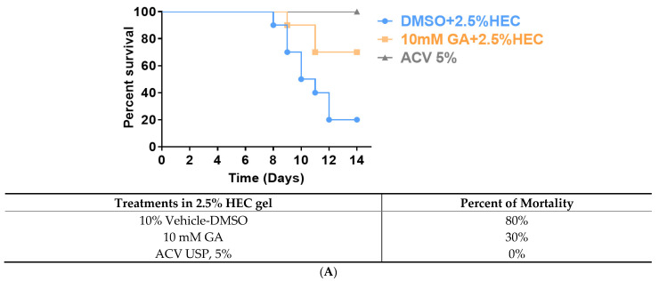
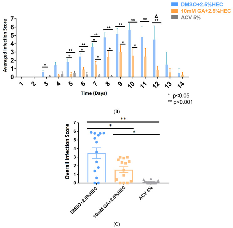
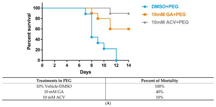
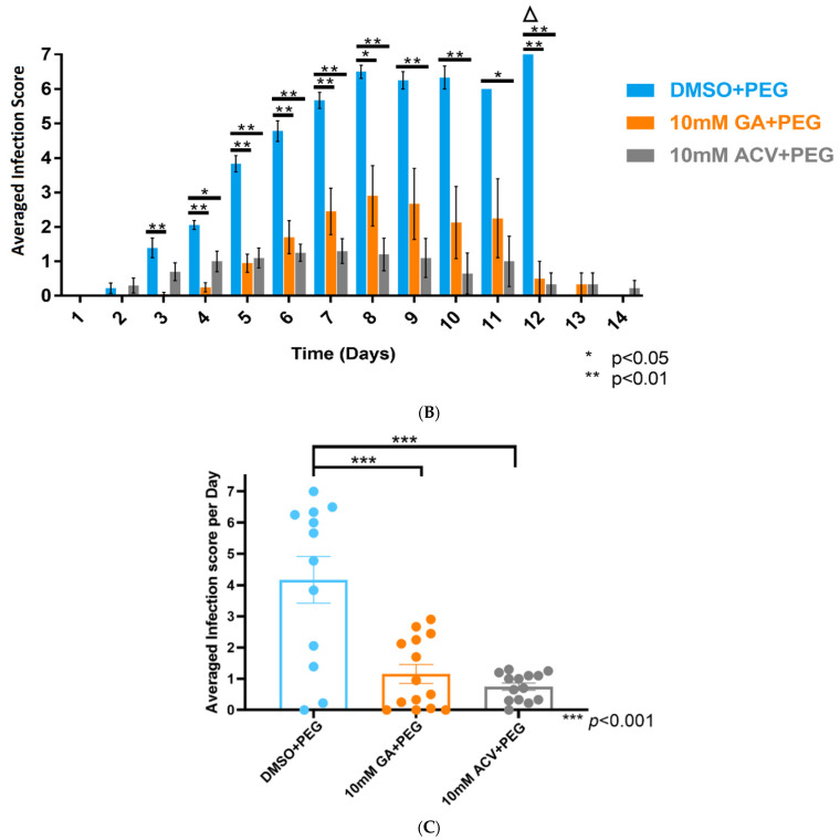
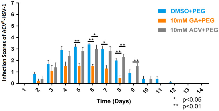
Herpes simplex virus type 1 (HSV-1) causes a lifelong latent infection with an estimated global prevalence of 66%. Primary and recurrent HSV infections are characterized by a tingling sensation, followed by an eruption of vesicles, which can cause painful erosions. Commonly used antiviral drugs against HSV infection are nucleoside analogues including acyclovir (ACV), famciclovir, and valacyclovir. Although these nucleoside analogues reduce morbidity and mortality in immunocompetent individuals, ACV-resistant HSV strains (ACVR-HSV) have been isolated from immunocompromised patients. Thus, ACVR-HSV infection poses a critical emerging public health concern. Recently, we reported that ginkgolic acid (GA) inhibits HSV-1 by disrupting viral structure, blocking fusion, and inhibiting viral protein synthesis. Additionally, we showed GA affords a broad spectrum of fusion inhibition of all three classes of fusion proteins, including those of HIV, Ebola, influenza A and Epstein Barr viruses. Here we report GA's antiviral activity against HSV-1 skin infection in BALB/cJ mice. GA-treated mice demonstrated a significantly reduced mortality rate and decreased infection scores compared to controls treated with dimethylsulfoxide (DMSO)-vehicle. Furthermore, GA efficiently inhibited ACVR-HSV-1 strain 17+ in vitro and in vivo. Since GA's mechanism of action includes virucidal activity and fusion inhibition, it is expected to work alone or synergistically with other anti-viral drugs, and we anticipate it to be effective against additional cutaneous and potentially systemic viral infections.

Keywords: acyclovir-resistance; antiviral; fusion inhibition; ginkgolic acid; herpes simplex type 1; virucidal activity; zosteriform infection.


Conflict of interest statement

The authors declare no conflict of interest.

Figures

Figure 1
Figure 1
Verification of acyclovir-resistant (ACVR) HSV-1 infection in Vero cells. (A) Flow chart of the methodology used to create ACV-resistant (ACVR) mutants, adapted from Sarisky et al. [33]. Verification of acyclovir-resistant (ACVR)-GFP-HSV-1 17+ in Vero cells. (B) Fluorescent images of 0.1 MOI GFP-HSV-1 infection in Vero cells with or without 45 µM ACV. FITC filter with excitation of 490 nm and emission of 525 nm was used to visualize viral plaques at 40× magnification; Scale bar represents 200 µm. (C) Fluorescent images of 0.1 MOI ACVR-GFP-HSV-1 infection in Vero cells with or without 45 µM ACV. FITC filter with excitation of 490 nm and emission of 525 nm was used to visualize viral plaques at 40× magnification. Scale bar represents 200 µm. (D) Plaque Assays of viral stocks (1 MOI) with or without 45 µM ACV in Vero cells in viral dilutions of 10−2 to 10−4, shown as qualitative image and in a table with averaged viral titers. (E) Western Blot analysis using antibodies directed against HSV-1 infected cellular protein 0 (ICP0; Santa-Cruz, sc-53070), glycoprotein D (gD; Santa-Cruz, sc-69802), thymidine kinase (TK; Invitrogen, PA5-67984), and GAPDH (0411; Santa-Cruz, sc-47724). M: Chameleon 800 Pre-stained Protein Ladder (Li-Cor, 928-80000). Signaling intensities of bands for each antibody have been normalized (as a percentage) to GFP-HSV-1. ACVR: ACVR-HSV-1. (F) Plaque Assays of GFP-HSV-1 17+ (0.1 MOI) and ACVR- HSV-1 (0.1 MOI) with various treatments in Vero cells such as DMSO (1:2000), 10 µM GA, and 10 µM ACV in viral dilutions of 10−2 to 10−4, shown as qualitative image and in a table with averaged viral titers.
Figure 1
Figure 1
Verification of acyclovir-resistant (ACVR) HSV-1 infection in Vero cells. (A) Flow chart of the methodology used to create ACV-resistant (ACVR) mutants, adapted from Sarisky et al. [33]. Verification of acyclovir-resistant (ACVR)-GFP-HSV-1 17+ in Vero cells. (B) Fluorescent images of 0.1 MOI GFP-HSV-1 infection in Vero cells with or without 45 µM ACV. FITC filter with excitation of 490 nm and emission of 525 nm was used to visualize viral plaques at 40× magnification; Scale bar represents 200 µm. (C) Fluorescent images of 0.1 MOI ACVR-GFP-HSV-1 infection in Vero cells with or without 45 µM ACV. FITC filter with excitation of 490 nm and emission of 525 nm was used to visualize viral plaques at 40× magnification. Scale bar represents 200 µm. (D) Plaque Assays of viral stocks (1 MOI) with or without 45 µM ACV in Vero cells in viral dilutions of 10−2 to 10−4, shown as qualitative image and in a table with averaged viral titers. (E) Western Blot analysis using antibodies directed against HSV-1 infected cellular protein 0 (ICP0; Santa-Cruz, sc-53070), glycoprotein D (gD; Santa-Cruz, sc-69802), thymidine kinase (TK; Invitrogen, PA5-67984), and GAPDH (0411; Santa-Cruz, sc-47724). M: Chameleon 800 Pre-stained Protein Ladder (Li-Cor, 928-80000). Signaling intensities of bands for each antibody have been normalized (as a percentage) to GFP-HSV-1. ACVR: ACVR-HSV-1. (F) Plaque Assays of GFP-HSV-1 17+ (0.1 MOI) and ACVR- HSV-1 (0.1 MOI) with various treatments in Vero cells such as DMSO (1:2000), 10 µM GA, and 10 µM ACV in viral dilutions of 10−2 to 10−4, shown as qualitative image and in a table with averaged viral titers.
Figure 2
Figure 2
GFP-HSV-1 average zosteriform infection scores of BALB/cJ mice (N = 10/treatment), following the application of DMSO in 2.5% HydroxyEthyl Cellulose (HEC) gel, 10 mM GA in HEC gel, and ACV USP 5%. (A) Age-matched BALB/cJ mice were inoculated with 6 × 10⁴ PFU of GFP-HSV-1, given respective treatments, and monitored for survival for 14 days. (B) Averaged infection scores of surviving animals in each treatment group across 14 days. (C) Combined and averaged infection score of all animals per day for 14 days. GA-treated animals demonstrated a significant reduction in the appearance of vesicles and erosions compared to vehicle-treated control animals. (B) Student independent t-tests (2-tailed) and (C) One-way ANOVA [F(2, 39) = 12.31]; * p < 0.05, ** p < 0.001. Δ indicates that 8 out of 10 animals in the DMSO-treated group died by day 12 p.i. All error bars represent SEM.
Figure 2
Figure 2
GFP-HSV-1 average zosteriform infection scores of BALB/cJ mice (N = 10/treatment), following the application of DMSO in 2.5% HydroxyEthyl Cellulose (HEC) gel, 10 mM GA in HEC gel, and ACV USP 5%. (A) Age-matched BALB/cJ mice were inoculated with 6 × 10⁴ PFU of GFP-HSV-1, given respective treatments, and monitored for survival for 14 days. (B) Averaged infection scores of surviving animals in each treatment group across 14 days. (C) Combined and averaged infection score of all animals per day for 14 days. GA-treated animals demonstrated a significant reduction in the appearance of vesicles and erosions compared to vehicle-treated control animals. (B) Student independent t-tests (2-tailed) and (C) One-way ANOVA [F(2, 39) = 12.31]; * p < 0.05, ** p < 0.001. Δ indicates that 8 out of 10 animals in the DMSO-treated group died by day 12 p.i. All error bars represent SEM.
Figure 3
Figure 3
GFP-HSV-1 average zosteriform infection scores of BALB/cJ mice, following the application of 10% vehicle-DMSO in Polyethylene glycol (PEG) (N = 9/group), 10 mM GA in PEG (N = 10/group), and 10 mM Acyclovir in PEG (N = 10/group). (A) Age-matched BALB/cJ mice were inoculated with 6 × 10⁴ PFU of GFP-HSV-1, given respective treatments, and monitored for survival for 14 days. (B) Averaged infection scores of surviving animals in each treatment group across 14 days. (C) Combined infection score of all animals, in their respective groups, per day for 14 days. Student independent t-tests (2-tailed); * p < 0.05; ** p < 0.01; *** p < 0.001. Δ indicates that all DMSO-treated animals died on day 12. All error bars represent SEM.
Figure 3
Figure 3
GFP-HSV-1 average zosteriform infection scores of BALB/cJ mice, following the application of 10% vehicle-DMSO in Polyethylene glycol (PEG) (N = 9/group), 10 mM GA in PEG (N = 10/group), and 10 mM Acyclovir in PEG (N = 10/group). (A) Age-matched BALB/cJ mice were inoculated with 6 × 10⁴ PFU of GFP-HSV-1, given respective treatments, and monitored for survival for 14 days. (B) Averaged infection scores of surviving animals in each treatment group across 14 days. (C) Combined infection score of all animals, in their respective groups, per day for 14 days. Student independent t-tests (2-tailed); * p < 0.05; ** p < 0.01; *** p < 0.001. Δ indicates that all DMSO-treated animals died on day 12. All error bars represent SEM.
Figure 4
Figure 4
ACVR-HSV-1 average zosteriform infection scores of BALB/cJ mice, following the application of 10% DMSO in polyethylene glycol (PEG), 10 mM GA in PEG, and 10 mM ACV in PEG (N = 5/group). Age-matched BALB/cJ mice were inoculated with 2.0 × 10⁵ PFU of ACVR-HSV-1 using the epidermal scarification-zosteriform model. Each group received the respective treatments and were monitored for 14 days. The infection scores of surviving animals were averaged in each treatment group for each day. GA-treated animals demonstrated a significant reduction in the appearance of vesicles and erosions compared to vehicle-treated control animals. Student independent t-tests (2-tailed); * p < 0.05; ** p < 0.01. All error bars represent SEM.

Similar articles

Cited by

References

    1. CDC Prevalence of Herpes Simplex Virus Type 1 and Type 2 in Persons Aged 14–49: United States, 2015–2016 (Number 304; February 2018) [(accessed on 11 July 2020)]; Available online: https://www.cdc.gov/nchs/products/databriefs/db304.htm#:~:text=The%20pre....
    1. James C., Harfouche M., Welton N.J., Turner K.M., Abu-Raddad L.J., Gottlieb S.L., Looker K.J. Herpes simplex virus: Global infection prevalence and incidence estimates, 2016. Bull. World Health Organ. 2020;98:315–329. doi: 10.2471/BLT.19.237149. - DOI - PMC - PubMed
    1. Lundberg P., Ramakrishna C., Brown J., Tyszka J.M., Hamamura M., Hinton D.R., Kovats S., Nalcioglu O., Weinberg K., Openshaw H. The immune response to herpes simplex virus type 1 infection in susceptible mice is a major cause of central nervous system pathology resulting in fatal encephalitis. J. Virol. 2008;82:7078–7088. doi: 10.1128/JVI.00619-08. - DOI - PMC - PubMed
    1. Piret J., Désormeaux A., Gourde P., Juhász J., Bergeron M.G. Efficacies of topical formulations of foscarnet and acyclovir and of 5-percent acyclovir ointment (Zovirax) in a murine model of cutaneous herpes simplex virus type 1 infection. Antimicrob. Agents Chemother. 2000;44:30–38. doi: 10.1128/AAC.44.1.30-38.2000. - DOI - PMC - PubMed
    1. Van Lint A., Ayers M., Brooks A.G., Coles R.M., Heath W.R., Carbone F.R. Herpes Simplex Virus-Specific CD8 T Cells Can Clear Established Lytic Infections from Skin and Nerves and Can Partially Limit the Early Spread of Virus after Cutaneous Inoculation. J. Immunol. 2004;172:392–397. doi: 10.4049/jimmunol.172.1.392. - DOI - PubMed

LinkOut - more resources