

Assessment of Photo-Induced Cytotoxic Activity of Cachrys sicula and Cachrys libanotis Enriched-Coumarin Extracts against Human Melanoma Cells
- PMID: 33435579
- PMCID: PMC7826759
- DOI: 10.3390/plants10010123
Assessment of Photo-Induced Cytotoxic Activity of Cachrys sicula and Cachrys libanotis Enriched-Coumarin Extracts against Human Melanoma Cells
Abstract
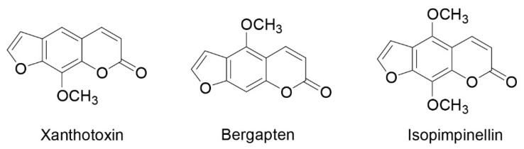
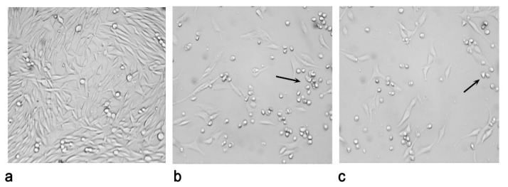
Photochemotherapy is one of the most interesting current therapeutic approaches for the treatment of melanoma. Different classes of naturally occurring phytochemicals demonstrated interesting photoactive properties. The aim of this study was to evaluate the photocytotoxic potential of two Cachrys species from Southern Italy: C. sicula and C. libanotis (Apiaceae). The enriched-coumarin extracts were obtained from aerial parts through both traditional maceration and pressurized cyclic solid-liquid (PCSL) extraction using Naviglio extractor®. Qualitative and quantitative analyses of furanocoumarins were performed with GC-MS. The photocytotoxic effects were verified on C32 melanoma cells irradiated at a dose of 1.08 J/cm2. The apoptotic responses were also assessed. Moreover, phenolic content and the in vitro antioxidant potential were estimated. Xanthotoxin, bergapten, and isopimpinellin were identified. All the samples induced concentration-dependent photocytotoxic effects (IC50 ranging from 3.16 to 18.18 μg/mL). The C. libanotis sample obtained with Naviglio extractor® was the most effective one (IC50 = 3.16 ± 0.21 μg/mL), followed by C. sicula sample obtained with the same technique (IC50 = 8.83 ± 0.20 μg/mL). Both Cachrys samples obtained through PCSL induced up-regulation of apoptotic signals such as BAX (Bcl2-associated X protein) and PARP (poly ADP-ribose polymerase) cleavage. Moreover, these samples proved to be more photoactive, giving a greater upregulation of p21 protein in the presence of UVA radiation. Obtained results suggest that investigated species could be promising candidates for further investigations aimed to find new potential drugs for the photochemotherapy of skin cancer.
Keywords: Apiaceae; Cachrys spp.; furanocoumarins; green extraction technology; photochemotherapy; skin cancer.
Conflict of interest statement
The authors declare no conflict of interest.
Figures


References
-
- Russo A., Cardile V., Graziano A.C.E., Avola R., Montenegro I., Cuellar M., Villena J., Madrid A. Antigrowth activity and induction of apoptosis in human melanoma cells by Drymis winteri forst extract and its active components. Chem. Biol. Interact. 2019;25:79–85. doi: 10.1016/j.cbi.2019.03.029. - DOI - PubMed
-
- De Oliveira Júnior R.G., Ferraz C.A.A., Silva M.G., de Lavor É.M., Rolim L.A., de Lima J.T., Fleury A., Picot L., de Souza Siqueira Quintans J., Quintans Junior L.J., et al. Flavonoids: Promising natural products for treatment of skin cancer (melanoma) In: Badria F.A., editor. Natural Products and Cancer Drug Discovery. InTech; Rijeka, Croatia: 2017. pp. 161–210.
LinkOut - more resources
Full Text Sources
Other Literature Sources
Research Materials
Miscellaneous
