

Centrosome-centric view of asymmetric stem cell division
- PMID: 33435817
- PMCID: PMC7881172
- DOI: 10.1098/rsob.200314
Centrosome-centric view of asymmetric stem cell division
Abstract
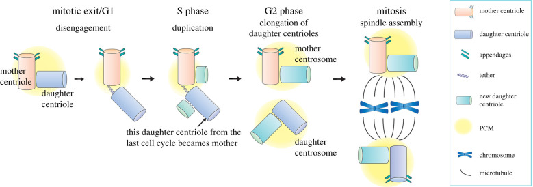
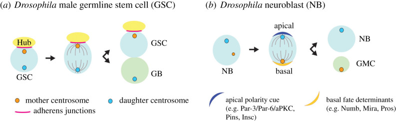
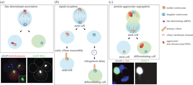
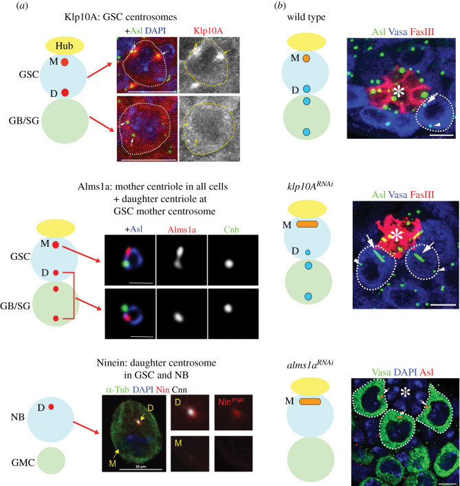
The centrosome is a unique organelle: the semi-conservative nature of its duplication generates an inherent asymmetry between 'mother' and 'daughter' centrosomes, which differ in their age. This asymmetry has captivated many cell biologists, but its meaning has remained enigmatic. In the last two decades, many stem cell types have been shown to display stereotypical inheritance of either the mother or daughter centrosome. These observations have led to speculation that the mother and daughter centrosomes bear distinct information, contributing to differential cell fates during asymmetric cell divisions. This review summarizes recent progress and discusses how centrosome asymmetry may promote asymmetric fates during stem cell divisions.
Keywords: asymmetric stem cell division; centrosomes; stem cell.
Conflict of interest statement
We declare we have no competing interests.
Figures
Similar articles
-
Centrosome asymmetry and inheritance during animal development.Curr Opin Cell Biol. 2012 Aug;24(4):541-6. doi: 10.1016/j.ceb.2012.05.005. Epub 2012 Jun 8. Curr Opin Cell Biol. 2012. PMID: 22683192 Free PMC article. Review.
-
Alstrom syndrome gene is a stem-cell-specific regulator of centriole duplication in the Drosophila testis.Elife. 2020 Sep 23;9:e59368. doi: 10.7554/eLife.59368. Elife. 2020. PMID: 32965218 Free PMC article.
-
Centrosome-Associated Degradation Limits β-Catenin Inheritance by Daughter Cells after Asymmetric Division.Curr Biol. 2015 Apr 20;25(8):1005-16. doi: 10.1016/j.cub.2015.02.020. Epub 2015 Mar 26. Curr Biol. 2015. PMID: 25819561
-
Regulation of centrosome movements by numb and the collapsin response mediator protein during Drosophila sensory progenitor asymmetric division.Development. 2013 Jul;140(13):2657-68. doi: 10.1242/dev.087338. Epub 2013 May 29. Development. 2013. PMID: 23720047
-
Fly meets yeast: checking the correct orientation of cell division.Trends Cell Biol. 2011 Sep;21(9):526-33. doi: 10.1016/j.tcb.2011.05.004. Epub 2011 Jun 24. Trends Cell Biol. 2011. PMID: 21705221 Free PMC article. Review.
Cited by
-
Asymmetry of centrosomes in Drosophila neural stem cells requires protein phosphatase 4.Mol Biol Cell. 2025 May 1;36(5):ar58. doi: 10.1091/mbc.E25-01-0021. Epub 2025 Mar 12. Mol Biol Cell. 2025. PMID: 40072519
-
Pericentrin interacts with Kinesin-1 to drive centriole motility.J Cell Biol. 2022 Sep 5;221(9):e202112097. doi: 10.1083/jcb.202112097. Epub 2022 Aug 5. J Cell Biol. 2022. PMID: 35929834 Free PMC article.
-
K-Ras Binds Calmodulin-Related Centrin1 with Potential Implications for K-Ras Driven Cancer Cell Stemness.Cancers (Basel). 2023 Jun 7;15(12):3087. doi: 10.3390/cancers15123087. Cancers (Basel). 2023. PMID: 37370697 Free PMC article.
-
Moonlighting at the Poles: Non-Canonical Functions of Centrosomes.Front Cell Dev Biol. 2022 Jul 14;10:930355. doi: 10.3389/fcell.2022.930355. eCollection 2022. Front Cell Dev Biol. 2022. PMID: 35912107 Free PMC article. Review.
-
APC/C-dependent degradation of Spd2 regulates centrosome asymmetry in Drosophila neural stem cells.EMBO Rep. 2023 Apr 5;24(4):e55607. doi: 10.15252/embr.202255607. Epub 2023 Feb 28. EMBO Rep. 2023. PMID: 36852890 Free PMC article.
References
-
- Boveri T 1887. Ueber den antheil des spermatozoon an der teilung des eies. Sitzungsber Ges Morph Physiol Much. 3, 151–164.
-
- Boveri T 1901. Zellenstudien IV. Uber die natur der centrosomen. Jena Zeitschr Naturw 35, 1–220.
-
- Boveri T 1914. Zur frage der entstehung maligner tumoren. Jena, Germany: Gustav Fischer Verlag.
Publication types
MeSH terms
Substances
LinkOut - more resources
Full Text Sources
Other Literature Sources
Medical
