. 2021 Jan 12;6(1):e00693-20.
doi: 10.1128/mSystems.00693-20.

Dinoroseobacter shibae Outer Membrane Vesicles Are Enriched for the Chromosome Dimer Resolution Site dif

Affiliations

Dinoroseobacter shibae Outer Membrane Vesicles Are Enriched for the Chromosome Dimer Resolution Site dif

Hui Wang et al. mSystems. .

Abstract

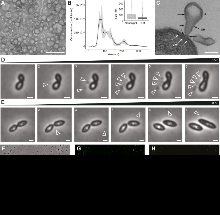
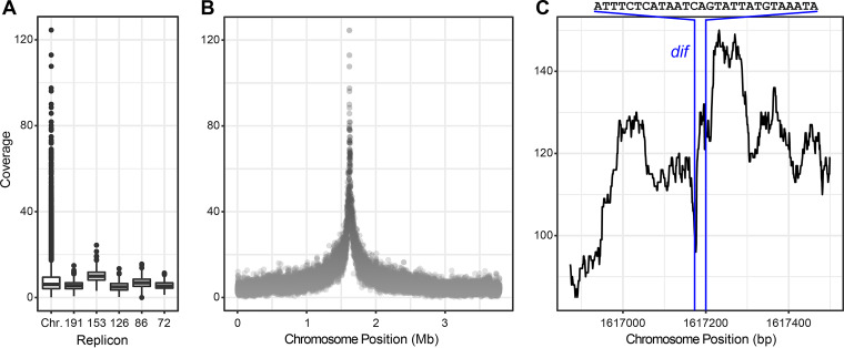
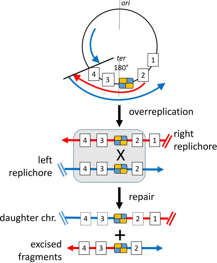
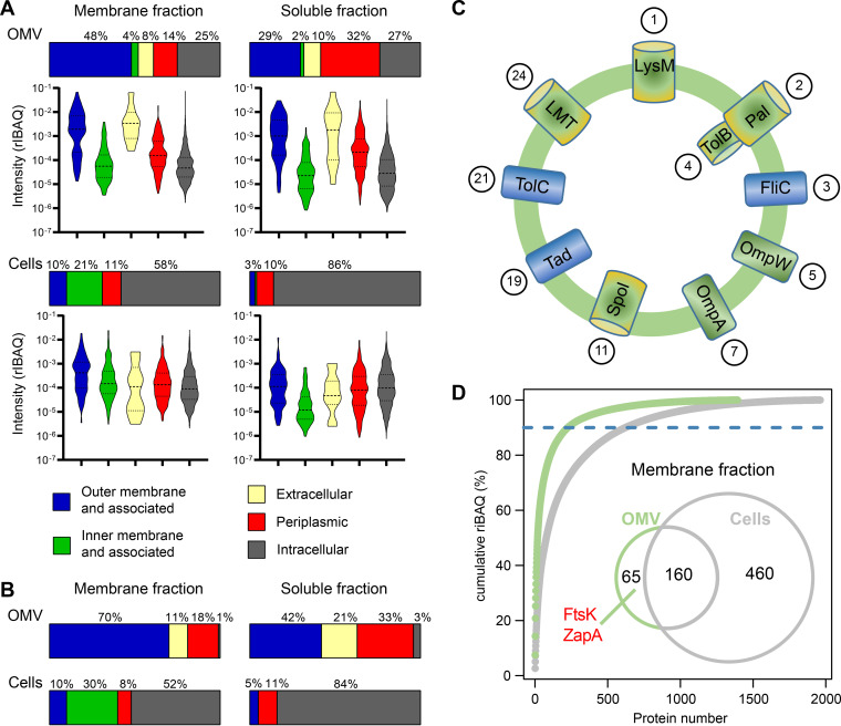
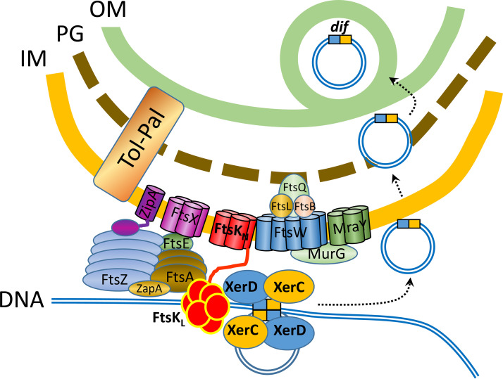
Outer membrane vesicles (OMVs) are universally produced by prokaryotes and play important roles in symbiotic and pathogenic interactions. They often contain DNA, but a mechanism for its incorporation is lacking. Here, we show that Dinoroseobacter shibae, a dinoflagellate symbiont, constitutively secretes OMVs containing DNA. Time-lapse microscopy captured instances of multiple OMV production at the septum during cell division. DNA from the vesicle lumen was up to 22-fold enriched for the region around the terminus of replication (ter). The peak of coverage was located at dif, a conserved 28-bp palindromic sequence required for binding of the site-specific tyrosine recombinases XerC/XerD. These enzymes are activated at the last stage of cell division immediately prior to septum formation when they are bound by the divisome protein FtsK. We suggest that overreplicated regions around the terminus have been repaired by the FtsK-dif-XerC/XerD molecular machinery. The vesicle proteome was clearly dominated by outer membrane and periplasmic proteins. Some of the most abundant vesicle membrane proteins were predicted to be required for direct interaction with peptidoglycan during cell division (LysM, Tol-Pal, Spol, lytic murein transglycosylase). OMVs were 15-fold enriched for the saturated fatty acid 16:00. We hypothesize that constitutive OMV secretion in D. shibae is coupled to cell division. The footprint of the FtsK-dif-XerC/XerD molecular machinery suggests a novel potentially highly conserved route for incorporation of DNA into OMVs. Clearing the division site from small DNA fragments might be an important function of vesicles produced during exponential growth under optimal conditions.IMPORTANCE Gram-negative bacteria continually form vesicles from their outer membrane (outer membrane vesicles [OMVs]) during normal growth. OMVs frequently contain DNA, and it is unclear how DNA can be shuffled from the cytoplasm to the OMVs. We studied OMV cargo in Dinoroseobacter shibae, a symbiont of dinoflagellates, using microscopy and a multi-omics approach. We found that vesicles formed during undisturbed exponential growth contain DNA which is enriched for genes around the replication terminus, specifically, the binding site for an enzyme complex that is activated at the last stage of cell division. We suggest that the enriched genes are the result of overreplication which is repaired by their excision and excretion via membrane vesicles to clear the divisome from waste DNA.

Keywords: DNA repair; OMV; circular chromosomes; replication termination; vesicles.


Figures

FIG 1
FIG 1
Vesicle formation in the D. shibae cell population. (A) Transmission electron microscopy (TEM) showing negatively stained vesicles purified by ultracentrifugation from an exponentially growing culture of D. shibae. (B) Size and abundance of vesicles determined by NanoSight particle tracking analysis. (Inset) Comparison of NanoSight and TEM quantification; n = 2,406 and 1,421, respectively. (C) TEM of ultrathin sections shows OMVs originating from the outer membrane. Scale bars in panels A and C, 200 nm. (D and E) Two instances of vesicle formation observed by phase-contrast time-lapse microscopy. Representative images are shown here, see also Movies S1 and S2 in the supplemental material. (D) Vesicle formation was followed over 13 h. An increasing number of OMVs were released from the division plane of the same cell over time (arrowheads). The vesicles stayed attached to the donor cell. The OMV-releasing cell stopped dividing during vesicle segregation. (E) Vesicle formation was followed over 8 h. A single OMV was formed and released into the supernatant. The OMV appeared at the division plane and started to move around the cell. Scale bars, 1 μm. (F to H) Light microscopic detection of DNA within OMVs. Phase contrast (F), membrane staining with FM1-43 (G), and DNA staining with DAPI (H) are shown. Scale bars in panels F to H, 10 μm.
FIG 2
FIG 2
DNA from OMVs of D. shibae was enriched for the region around the terminus of replication (ter). (A) Sequence coverage of chromosome and plasmids in DNA inside OMVs. Median, minimum, and maximum values are shown. Plasmids are abbreviated according to their size: 191, 152, 126, 86 and 72 kb. See Fig. S3 for two additional biological replicates. (B) Sequence coverage of the chromosome showing overrepresentation of the region on both sides of position ∼1.6 Mb, which is the region around the replication terminus. Coverage was calculated for a sliding window of 500 nt in panels A and B. See Data Set S1, sheet 3, for an identification of the enriched genes at coverages of >40-fold and >100-fold. (C) Sequence coverage of the region around chromosome position 1.6 Mb at single-base resolution. The position and 28-bp nucleotide sequence of dif are indicated.
FIG 3
FIG 3
Working hypothesis for enrichment of dif-containing DNA in OMVs of D. shibae. The chromosome is schematically shown by a black circle. Origin of replication (ori) and terminus region (ter) at 180° are indicated. The palindromic binding sequence dif is represented by a blue and yellow box, and genes in the terminus region are shown as boxed numbers 1 to 4. Left and right replichores are represented by blue and red lines, respectively. Overreplicated genes are boxed. Arrows represent the replication fork. Only the leading strand of each replichore is shown. chr, chromosome. Two possible mechanisms of repair of overreplicated regions are shown in detail in Fig. S4.
FIG 4
FIG 4
Vesicles of D. shibae are enriched for outer membrane and periplasmic proteins, of which many are related to cell division. Predicted localization of all detected (A) and the 30 most abundant (B) soluble and membrane proteins from D. shibae vesicles and cells. (A) Violin plots show the frequency distribution of relative iBAQ values within the groups of each fraction. Median and quantile borders are indicated by dashed and dotted lines, respectively. In total, 1,393 proteins were identified in the membrane fraction of vesicles, 2,223 in the soluble fraction of vesicles, 1,962 proteins in the membrane fraction of cells, and 2,548 proteins in the soluble fraction of cells. (C) Scheme of the most abundant proteins in the OMV membrane. Proteins are ordered in clockwise fashion according to their relative abundance (their rank among the top 30 most abundant vesicle membrane proteins is indicated in circles). All of the predicted functions are hypothetical. LysM, a protein containing a LysM domain required for peptidoglycan hydrolysis; Pal, an outer membrane lipoprotein preferentially located at the septum; TolB, the periplasmic component of the Pal-Tol complex required for cell division; FliC, flagellum filament protein; OmpW, an abundant pore in the outer membrane; OmpA, one of the most abundant outer membrane proteins with two domains, a pore, and a cytoplasmic domain that interacts with peptidoglycan; Spo1, a protein containing a SPOR domain which preferentially binds denuded peptidoglycan at the septum; Tad, part of the Tad (tight adherence) pilus assembly and secretion system common in roseobacters; TolC, the outer membrane component of an energy driven multidrug efflux pump in Gram-negative bacteria; LMT, a lytic murein transglycosylase that divides the septal murein into separate layers. See Results and Discussion for details on those proteins. (D) Abundant OMV membrane proteins. Venn diagram shows unique and overlapping number of proteins comprising 90% of cumulative riBAQ of the respective protein fraction in cells and OMVs. ZapA and FtsK within the OMV fraction are indicated.
FIG 5
FIG 5
Scheme of the FtsK-dif-XerC/XerD protein complex in the divisome and the export of DNA into OMVs. A subset of the proteins comprising the divisome and their localization at the cell envelope is schematically shown. FtsKN is the N-terminal domain of FtsK, and FtsKL, is its C-terminal domain. FtsK is a DNA translocase that moves the replichore toward dif and activates the site-specific recombinases XerC/XerD. We hypothesize that these enzymes excise overreplicated genes around the terminus which then enter the periplasm and are enclosed by the outer membrane. It is unknown if the excised DNA is circular or linear. OM, outer membrane; PG, peptidoglycan; IM, inner membrane. Modified from references and with incorporation of information from references , and .

Similar articles

Cited by

References

    1. Deatherage BL, Cookson BT. 2012. Membrane vesicle release in bacteria, eukaryotes, and archaea: a conserved yet underappreciated aspect of microbial life. Infect Immun 80:1948–1957. doi:10.1128/IAI.06014-11. - DOI - PMC - PubMed
    1. Gould SB, Garg SG, Martin WF. 2016. Bacterial vesicle secretion and the evolutionary origin of the eukaryotic endomembrane system. Trends Microbiol 24:525–534. doi:10.1016/j.tim.2016.03.005. - DOI - PubMed
    1. Kulp A, Kuehn MJ. 2010. Biological functions and biogenesis of secreted bacterial outer membrane vesicles. Annu Rev Microbiol 64:163–184. doi:10.1146/annurev.micro.091208.073413. - DOI - PMC - PubMed
    1. Schwechheimer C, Kuehn MJ. 2015. Outer-membrane vesicles from Gram-negative bacteria: biogenesis and functions. Nat Rev Microbiol 13:605–619. doi:10.1038/nrmicro3525. - DOI - PMC - PubMed
    1. Orench-Rivera N, Kuehn MJ. 2016. Environmentally controlled bacterial vesicle-mediated export. Cell Microbiol 18:1525–1536. doi:10.1111/cmi.12676. - DOI - PMC - PubMed

LinkOut - more resources