. 2021 Jan 12;12(1):77.
doi: 10.1038/s41419-020-03372-2.

SIRT6 promotes angiogenesis and hemorrhage of carotid plaque via regulating HIF-1α and reactive oxygen species

Affiliations

SIRT6 promotes angiogenesis and hemorrhage of carotid plaque via regulating HIF-1α and reactive oxygen species

Zhou Yang et al. Cell Death Dis. .

Abstract

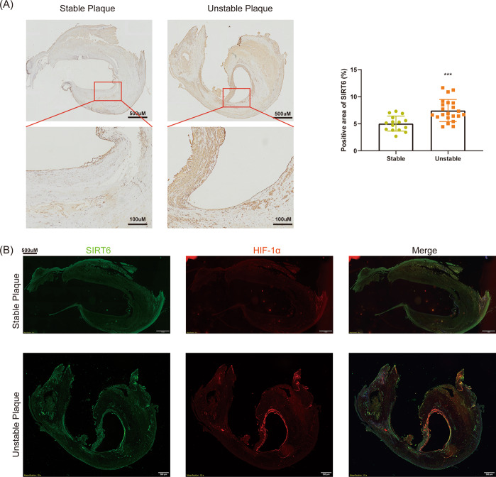
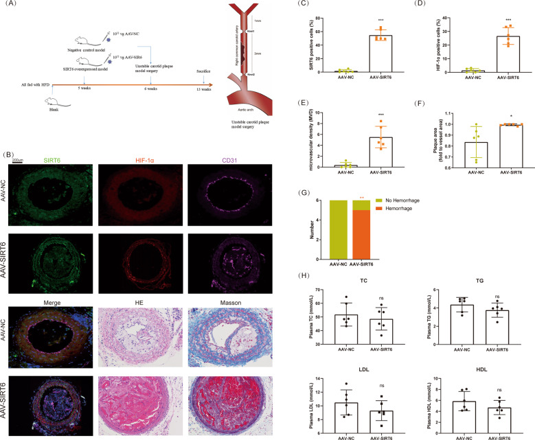
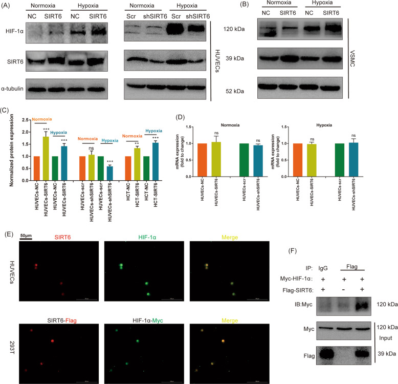
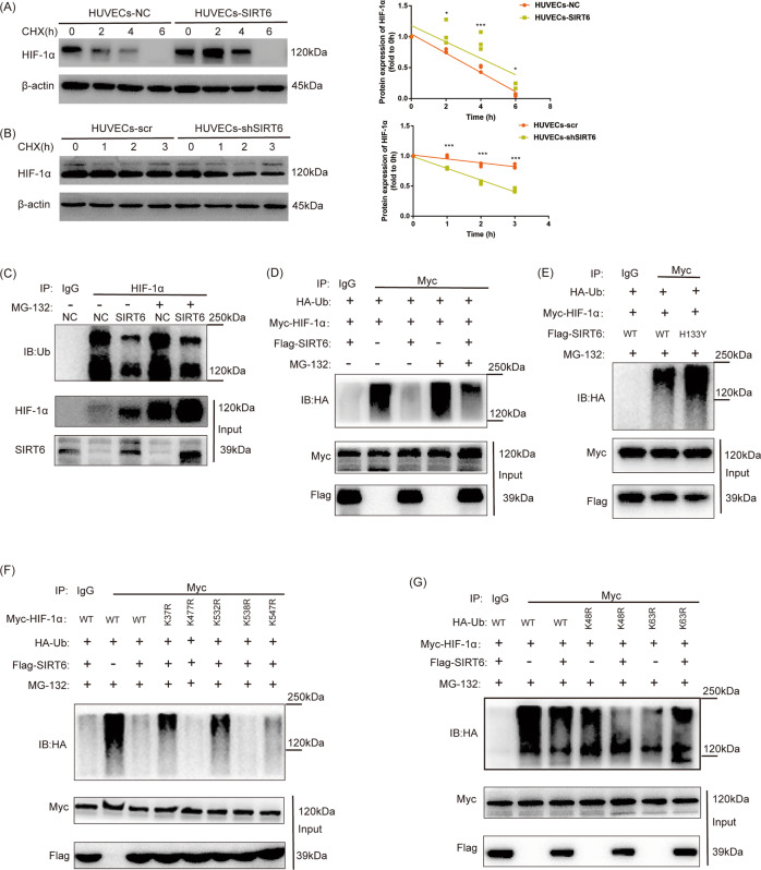
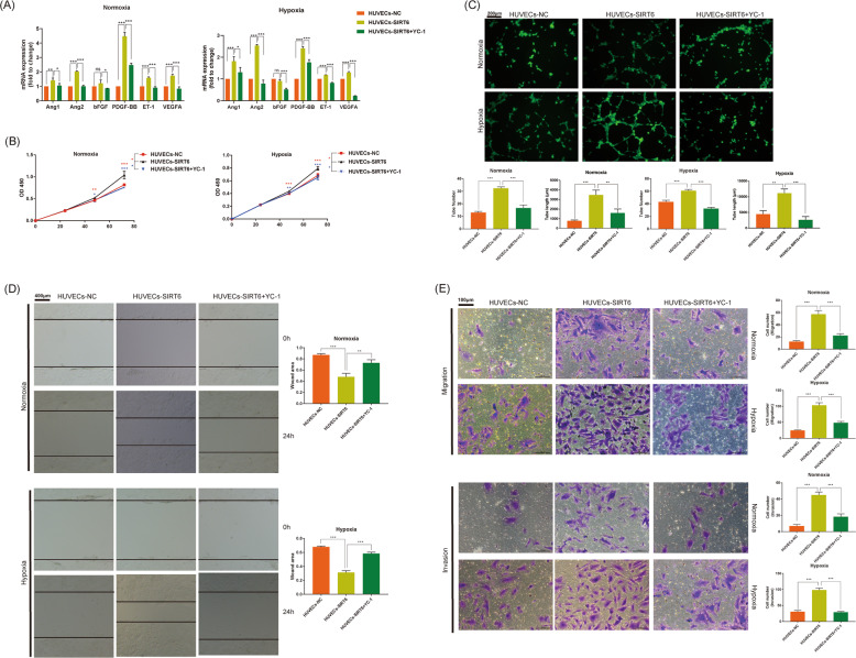
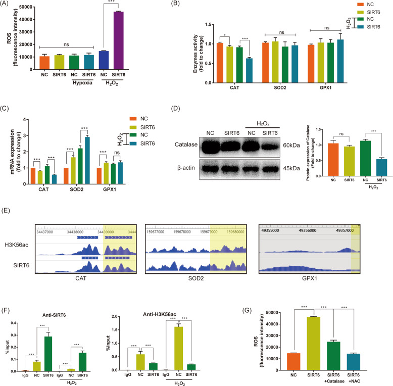
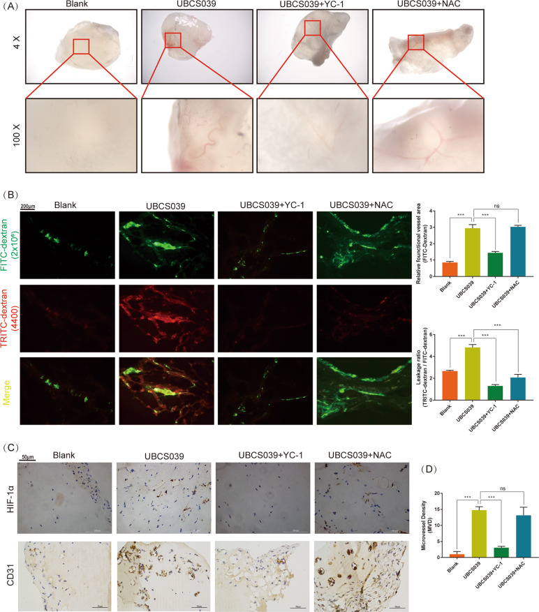
As a member of Sirtuins family, SIRT6 participates in the physiological and pathological progress of DNA repair, anti-aging, metabolism, and so on. Several studies have demonstrated that knockdown of SIRT6 inhibited the development of atherosclerosis (AS), indicated SIRT6 as a protective factor for AS. However, we confirmed SIRT6 was significantly overexpressed in human unstable carotid plaques compared with stable carotid plaques. This result indicated a more complex role of SIRT6 in AS. Furthermore, we constructed mice model with unstable carotid plaque and injected them with SIRT6 overexpressed adeno-associated virus (AAV-SIRT6). AAV-SIRT6 significantly promoted angiogenesis as well as hemorrhage in plaques. In vitro, we demonstrated overexpression of SIRT6 prevented HIF-1α from degradation by deubiquitination at K37 and K532 of HIF-1α, thus promoted the expression of HIF-1α under both normoxia and hypoxia in human umbilical vein endothelial cells (HUVECs). Through regulating HIF-1α, overexpression of SIRT6 promoted invasion, migration, proliferation, as well as tube formation ability of HUVECs. Interestingly, under different conditions, SIRT6 played different roles in the function of HUVECs. Under oxidative stress, another important pathological environment for AS, SIRT6 bound to the promoter of Catalase, a main reactive oxygen species scavenger, and depleted H3K56 acetylation, thus inhibited expression and activity of Catalase at the transcriptional level. Subsequently, inhibited Catalase promoted reactive oxygen species (ROS) under oxidative stress. Accumulated ROS further aggravated oxidative stress injury of HUVECs. On one hand, SIRT6 promoted angiogenesis in plaque via HIF-1α under hypoxia. On the other hand, SIRT6 promoted injury of neovascular via ROS under oxidative stress. It is this process of continuous growth and damage that leads to hemorrhage in carotid plaque. In conclusion, we innovatively confirmed SIRT6 promoted the angiogenesis and IPH via promoting HIF-1α and ROS in different environments, thus disclosed the unknowing danger of SIRT6.


Conflict of interest statement

The authors declare that they have no conflict of interest.

Figures

Fig. 1
Fig. 1. SIRT6 was overexpressed in unstable carotid plaque accompanied by co-location of HIF-1α.
A IHC of SIRT6 in stable (n = 14) and unstable carotid plaques (n = 22). B Co-location of SIRT6 and HIF-1α in stable and unstable carotid plaques (***P < 0.001).
Fig. 2
Fig. 2. Overexpression of SIRT6 promoted angiogenesis and hemorrhage in carotid plaque.
A Establishment of a mice model with unstable carotid plaque. SIRT6 was overexpressed by adenovirus associated virus (n = 6). B Immunofluorescence of SIRT6, HIF-1α, and CD31; HE staining; Masson staining of carotid plaque in mice model. C Analysis of the expression of SIRT6 in carotid plaque. D Analysis of the expression of HIF-1α in carotid plaque. E Microvascular density (MVD, marked by CD31) in carotid plaque. F, G Plaque area and hemorrhage analyzed by HE staining and Masson staining. H Quantification of total cholesterol (TC), triglyceride (TG), low-density lipoprotein (LDL), and high-density lipoprotein (HDL) in serum of mouse model (ns: no significance, *P < 0.05, **P < 0.01, ***P < 0.001).
Fig. 3
Fig. 3. SIRT6 promoted the expression of HIF-1α in HUVECs under both normoxia and hypoxia.
A The expression of HIF-1α under hypoxia (CoCl2) and normoxia after overexpression and downregulation of SIRT6 in HUVECs. B The expression of HIF-1α under hypoxia (CoCl2) and normoxia after overexpression of SIRT6 in VSMC. C Quantitative densitometric analysis of HIF-1α levels in panels A and B. D The mRNA expression of HIF-1α under hypoxia and normoxia after overexpression and downregulation of SIRT6 in HUVECs performed by RT-qPCR. E Colocalization of HIF-1α (Myc-tag) and SIRT6 (Flag-tag) under hypoxia in HUVECs and 293T performed by IF (magnification: ×200). F Co-IP of HIF-1α-Myc and SIRT6-Flag under hypoxia in 293T cell (ns: no significance, ***P < 0.001).
Fig. 4
Fig. 4. Overexpression of SIRT6 inhibited ubiquitin-proteasome degradation of HIF-1α.
(C, D, E, F, G: Detection of ubiquitination of HIF-1α performed by Co-IP under hypoxia). A Overexpression of SIRT6 prevented the degradation of HIF-1α under hypoxia after the treatment of CHX (20 µg/ml for 0–6 h). B Downregulation of SIRT6 promoted the degradation of HIF-1α under hypoxia after the treatment of CHX (20 µg/ml for 0–3 h). C Ubiquitination of HIF-1α in HUVECs-NC and HUVECs-SIRT6 with or without proteasome inhibitor MG-132 treatment (10 μm, 6 h). D SIRT6 promoted deubiquitination of HIF-1α. HUVECs were transfected with HA-Ub, Myc-HIF-1α, and SIRT6-Flag, respectively. E Inhibition of histone deacetylase activity of SIRT6 inhibited its deubiquitination effect at HIF-1α. HUVECs were transfected with HA-Ub, Myc-HIF-1α, and SIRT6-Flag (wild type: WT or histone deacetylase activity inhibited mutant: H133Y), respectively. F K37 and K532 are the main SIRT6 deubiquitination sites of HIF-1α. HUVECs was transfected with HA-Ub, SIRT6-Flag, and different Myc-HIF-1α mutant (K37R/K477R/K532R/K538R/K547R). G SIRT6 promoted the deubiquitination of HIF-1α via Ubiquitin-K63. HUVECs were transfected with SIRT6-Flag, Myc-HIF-1α, and HA-Ub (WT, K48R, and K63R mutant) (*P < 0.05, ***P < 0.001).
Fig. 5
Fig. 5. SIRT6 promoted migration, proliferation, and tube formation ability of HUVECs via regulation of HIF-1α.
A The expression of HIF-1α regulated angiogenic genes. B Cell proliferation of HUVECs-NC, HUVECs-SIRT6, and HUVECs-SIRT6 treated with HIF-1α inhibitor YC-1 (1 μm for 0–72 h) under both hypoxia and normoxia, performed by CCK8 assays. C Tube formation ability of HUVECs-NC, HUVECs-SIRT6, and HUVECs-SIRT6 pre-treated with HIF-1α inhibitor YC-1 (1 μm for 24 h) under both hypoxia and normoxia (magnification: ×200). D Invasion and migration ability of HUVECs-NC, HUVECs-SIRT6, and HUVECs-SIRT6 treated with HIF-1α inhibitor YC-1 (1 μm for 24 h) under both hypoxia and normoxia, performed by transwell assays (magnification: ×400). E Migration ability of HUVECs-NC, HUVECs-SIRT6, and HUVECs-SIRT6 treated with HIF-1α inhibitor YC-1 (1 μm for 24 h) under both hypoxia and normoxia, performed by wound healing assay (magnification: ×100) (ns: no significance, *P < 0.05, **P < 0.01, ***P < 0.001).
Fig. 6
Fig. 6. SIRT6 promoted reactive oxygen species via H3K56ac of Catalase in HUVECs under oxidative stress.
A ROS of HUVECs-NC and HUVECs-SIRT6 under normoxia (normal group), hypoxia, and oxidative stress (treated with 1 mM H2O2 for 3 h). B The enzyme activity of three main ROS scavengers: Catalase (CAT), superoxide dismutase (SOD2), and glutathione peroxidase (GPX1), with or without H2O2 treatment. C The mRNA expression of CAT, SOD2, and GPX1, with or without H2O2 treatment. D Protein expression of Catalase with or without H2O2 treatment performed by western blotting. E SIRT6 and H3K56ac enrichment at the promoter of CAT, SOD2, and GPX1 detected in CHIP-sequence datasets GSE102813. F CHIP analysis of HUVECs-NC and HUVECs-SIRT6 using IgG, anti-SIRT6, and anti-H3K56ac, respectively. G Transfection of pcDNA3.1-Catalase and treatment of N-acetyl-L-cysteine (NAC, 5 mM for 3 h) rescued increased ROS in HUVECs-SIRT6 (ns: no significance, **P < 0.01, ***P < 0.001).
Fig. 7
Fig. 7. SIRT6 promoted angiogenesis as well as vessel permeability in vivo (N = 5, each group).
A Matrigel plugs deprived from mice after 14 days of subcutaneous implantation. UBCS039: SIRT6 activator, 100 μm; YC-1: HIF-1α inhibitor, 2 μm; NAC: ROS scavenger, 10 μm. All drugs were blended with Matrigel gel before implantation. B FITC-dextran (2 × 106 MW) accompanied by TRITC-dextran (4400 MW) was injected before the scarification of mice. FITC-dextran (2 × 106 MW): functional vessel, TRITC-dextran (4400 MW): leaky blood cells from vessels. TRITC-dextran/FITC-dextran area (leakage ratio): vessel permeability (magnification: ×100). C Expression of HIF-1α and microvascular density (MVD, marked by CD31) of all matrigel plugs performed by IHC (magnification: ×400) (ns: no significance, ***P < 0.001).

References

    1. Os, H. J. A. V., Mulder, I. A., Broersen, A., Algra, A. & Wermer, M. J. H. Migraine and cerebrovascular atherosclerosis in patients with ischemic stroke. Stroke48, 1973–1975 (2017). - PubMed
    1. Loftus, I. M. Intraplaque Hemorrhage and Progression of Coronary Atheroma. N. Engl. J. Med. 349, 2316–2325 (2014). - PubMed
    1. Koole D, Heyligers J, Moll FL, Pasterkamp G. Intraplaque neovascularization and hemorrhage: markers for cardiovascular risk stratification and therapeutic monitoring. J. Cardiovascular Med. 2012;13:635–639. doi: 10.2459/JCM.0b013e3283590cd2. - DOI - PubMed
    1. Michishita E, et al. SIRT6 is a histone H3 lysine 9 deacetylase that modulates telomeric chromatin. Nature. 2008;452:492–496. doi: 10.1038/nature06736. - DOI - PMC - PubMed
    1. Jain AK, et al. LncPRESS1 Is a p53-Regulated LncRNA that Safeguards Pluripotency by Disrupting SIRT6-Mediated De-acetylation of Histone H3K56. Mol. Cell. 2016;64:967–981. doi: 10.1016/j.molcel.2016.10.039. - DOI - PMC - PubMed

Publication types

LinkOut - more resources