Review
. 2021 Jan 12;12(1):71.
doi: 10.1038/s41419-020-03314-y.

Structure, function, and pathology of protein O-glucosyltransferases

Affiliations
Review

Structure, function, and pathology of protein O-glucosyltransferases

Muhammad Zubair Mehboob et al. Cell Death Dis. .

Abstract

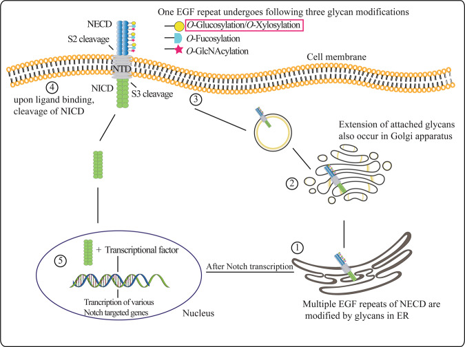
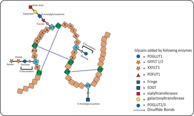
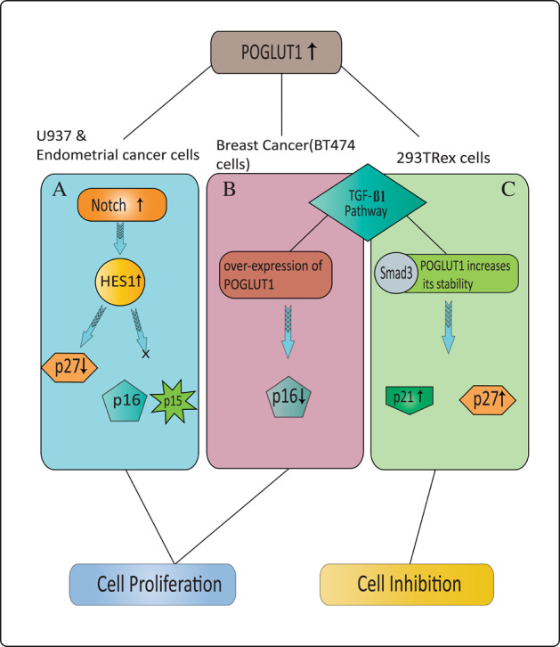
Protein O-glucosylation is a crucial form of O-glycosylation, which involves glucose (Glc) addition to a serine residue within a consensus sequence of epidermal growth factor epidermal growth factor (EGF)-like repeats found in several proteins, including Notch. Glc provides stability to EGF-like repeats, is required for S2 cleavage of Notch, and serves to regulate the trafficking of Notch, crumbs2, and Eyes shut proteins to the cell surface. Genetic and biochemical studies have shown a link between aberrant protein O-glucosylation and human diseases. The main players of protein O-glucosylation, protein O-glucosyltransferases (POGLUTs), use uridine diphosphate (UDP)-Glc as a substrate to modify EGF repeats and reside in the endoplasmic reticulum via C-terminal KDEL-like signals. In addition to O-glucosylation activity, POGLUTs can also perform protein O-xylosylation function, i.e., adding xylose (Xyl) from UDP-Xyl; however, both activities rely on residues of EGF repeats, active-site conformations of POGLUTs and sugar substrate concentrations in the ER. Impaired expression of POGLUTs has been associated with initiation and progression of human diseases such as limb-girdle muscular dystrophy, Dowling-Degos disease 4, acute myeloid leukemia, and hepatocytes and pancreatic dysfunction. POGLUTs have been found to alter the expression of cyclin-dependent kinase inhibitors (CDKIs), by affecting Notch or transforming growth factor-β1 signaling, and cause cell proliferation inhibition or induction depending on the particular cell types, which characterizes POGLUT's cell-dependent dual role. Except for a few downstream elements, the precise mechanisms whereby aberrant protein O-glucosylation causes diseases are largely unknown, leaving behind many questions that need to be addressed. This systemic review comprehensively covers literature to understand the O-glucosyltransferases with a focus on POGLUT1 structure and function, and their role in health and diseases. Moreover, this study also raises unanswered issues for future research in cancer biology, cell communications, muscular diseases, etc.


Conflict of interest statement

The authors declare that they have no conflict of interests.

Figures

Fig. 1
Fig. 1. Cellular regulation and representation of glycans.
Depending on the intracellular compartments, mechanisms altering the intracellular location of glycosyltransferases and glycosidases can influence and regulate glycans modfication. A Secretory glycosylation occurs mainly in the ER and Golgi apparatus for secreted or transmembrane proteins where glycosyltransferases add and extend glycan modification in an ordered sequential manner. Cellular receptors and secretory proteins are transported to the cell surface and extracellular space, respectively. B Intracellular glycosylation involves the addition of glycans by OGT to proteins that reside mainly in the cytoplasm and nucleus. This modification of a Ser/Thr residue has a short lifetime and is cleaved by OGA, providing a target site for the second same modification or phosphorylation of Ser/Thr.
Fig. 2
Fig. 2. A representation of Notch structure, modification, and signaling.
Notch receptor involved in several biological processes has three domains: NECD, NTD, and NICD. The NECD comprises a set of EGF-like repeats which undergo glycosylation. (1) The properly folded EGF repeats of NECD are modified with glycans added by glycan-specific enzymes and (2) these glycans are further extended in the Golgi apparatus or even also in the ER. (3) Sugar decorated Notch is transported to the cell membrane through membrane-bound vesicles. (4) After ligand (Delta, Jagged) binding, the ADAM proteases and γ-secretases execute Notch S2 and S3 cleavage, respectively, ultimately releasing the NICD that moves to the nucleus (5) where it binds the Notch pathway effector proteins to induce the expression of Notch target genes.
Fig. 3
Fig. 3. A comparison of structural domains of POGLUTs including Rumi.
A O-glucosyltransferases have a signal peptide (SP) at the beginning of sequence, a CAP10 domain, and a ER-retention signal. Both KTEL and REEL retention signals have a lower affinity for ERD receptors than the KDEL signal. POGLUT1 shares 52% identity with Rumi and is the only enzyme that can rescue O-glucosylation function in rumi mutant flies. Although POGLUT2 and 3 show O-glucosyltransferase activity, but both enzymes are unable to rescue Rumi loss-of-function phenotype and only share 24% and 20.3% identity with POGLUT1, respectively, indicating different target sequence for modification. B Multiple sequence alignment of glucosyltransferases indicates the presence of DXD-like motifs, whose mutation can abolish O-glucosylation activity of enzymes. Moreover, POGLUT 2/3 lacks the WEGG motif considered necessary for UDP-glucose binding.
Fig. 4
Fig. 4. EGF-like repeat in the NECD domain and its modifications.
Multiple EGF-like repeats present in the NECD harbor consensus sequences having a target site for glycosyltransferases. POGLUT1 adds O-Glc to a Ser residue in the consensus sequence C1-X-S-X-P/A-C2, which can further be extended to Xyl-Xyl-Glc-O trisaccharide by GXYLT1/2 and XXYLT1. The serine between C1 and C2 is highly conserved relative to other glycosylated sites, where both Ser and Thr can be found, and its the conformation of C1–C2 motif because of which POGLUTs can not glucosylate the Thr residue. POGLUT2 and 3 glucosylate a Ser residue conserved in C3–C4 of the EGF-like repeats and its further extension is not yet reported. POFUT1 adds O-Fuc to Ser/Thr residues between C2 and C3 of EGF repeats, which can further be elongated by Fringe enzyme. Moreover, the Ser/Thr residues between C5 and C6 is O-GlcNAcylated by a EOGT in Drosophila and mammals.
Fig. 5
Fig. 5. POGLUT1 mutations associated to cause human diseases.
Several mutations of POGLUT1 have been implicated in human DDD4/GGD and LGMDR21. So far, only two nonsense mutations are reported in a signal peptide of POGLUT1 in DDD4/GGD patients. Most of the missense, nonsense, and frameshift variants of POGLUT1 are reported in the CAP10 catalytic domain, which harbors many essential amino acids required for O-glucosylation activity. A single change of essential residues of CAP10 domain can abolish O-glucosyltransferase activity as previously observed. The nonsense and frameshift mutations are resulted either in mRNA decay or the formation of a truncated POGLUT1 leading to DDD4/GGD and LGMDR21 disease.
Fig. 6
Fig. 6. Partial signaling pathways reported based on specific cell types by POGLUT1 for cell proliferation and inhibition.
Dual role of POGLUT1 in cell cycle progression or supression is cell-type specific. Up to now, a complete signaling pathway for cell proliferation or inhibition in response to overexpression of POGLUT1 is not fully explained; however, POGLUT1’ association with Notch and TGF-β1 signaling pathways has been observed. A In U937 and endometrial cancer cells, higher expression of POGLUT1 increases Notch signaling and HES-1 level, which in turn decreases P27 level without altering p15 and p16, and induces cell cycle progression or tumorigenesis. B Evidently, decreased expression level of p16 upon overexpressing POGLUT1 has also been observed, which explains the involvement of more than one signaling route in regulating CDKIs level. Upregulated POGLUT1 reduces Smad3 phosphorylation in breast cancer cells by repressing p16 expression in the presence of TGF-β1, thus promoting BT474 cell proliferation. C In contrast, overexpression of POGLUT1 in 293TRex cells triggers cell cycle arrest and increases Smad3 protein stability via inhibiting its proteasomal degradation. In fact, POGLUT1 enhances TGF-β1 signaling by modulating Smad3 expression and increases level of CDKIs p27 and p21, which bind with CDKs enzymes to block cell proliferation.

Similar articles

Cited by

References

    1. Reily, C., Stewart, T. J., Renfrow, M. B. & Novak, J. Glycosylation in health and disease. Nat. Rev. Neurol. 15, 346–366 (2019). - PMC - PubMed
    1. Zachara, N., Akimoto, Y. & Hart, G. W. in Essentials of Glycobiology. 3rd edn (Cold Sping Harbor, NY, 2017).
    1. Stanley P. Golgi glycosylation. Cold Spring Harb. Perspect. Biol. 2011;3:a005199. doi: 10.1101/cshperspect.a005199. - DOI - PMC - PubMed
    1. Hart, G. W. & Akimoto, Y. in Essentials of Glycobiology. 2nd edn (Cold Sping Harbor, NY, 2009).
    1. Moremen KW, Tiemeyer M, Nairn AV. Vertebrate protein glycosylation: diversity, synthesis and function. Nat. Rev. Mol. Cell Biol. 2012;13:448–462. doi: 10.1038/nrm3383. - DOI - PMC - PubMed

Publication types

Substances

LinkOut - more resources