. 2021 Jan 12;12(1):312.
doi: 10.1038/s41467-020-20588-0.

Carbon nano-onion-mediated dual targeting of P-selectin and P-glycoprotein to overcome cancer drug resistance

Affiliations

Carbon nano-onion-mediated dual targeting of P-selectin and P-glycoprotein to overcome cancer drug resistance

Hai Wang et al. Nat Commun. .

Abstract

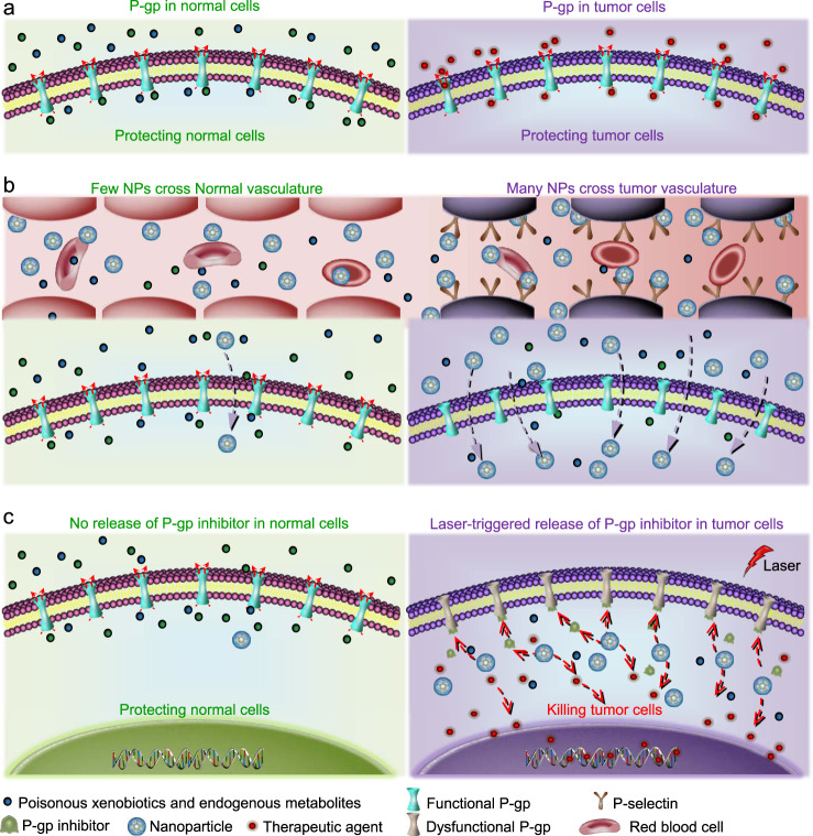
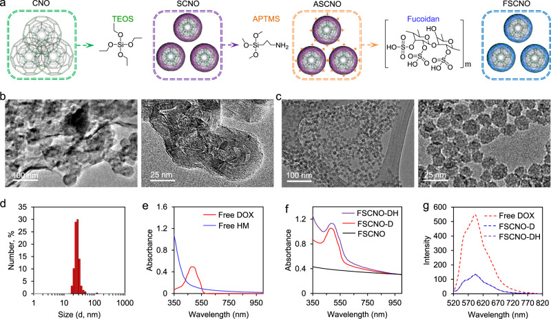
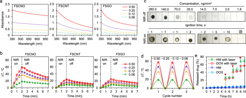
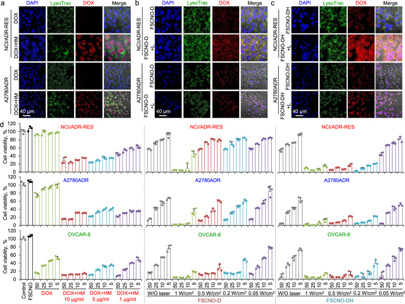
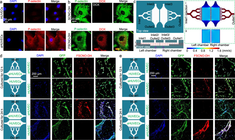
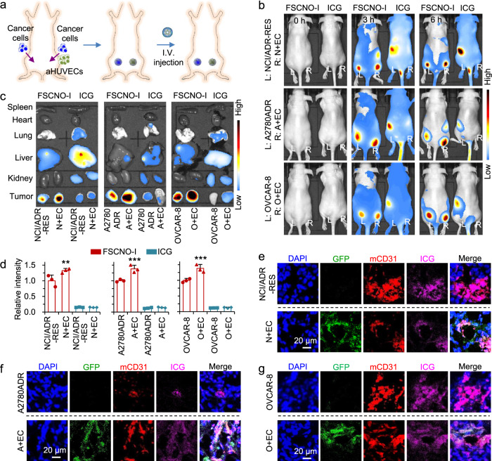
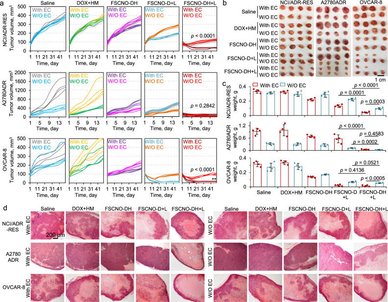
The transmembrane P-glycoprotein (P-gp) pumps that efflux drugs are a major mechanism of cancer drug resistance. They are also important in protecting normal tissue cells from poisonous xenobiotics and endogenous metabolites. Here, we report a fucoidan-decorated silica-carbon nano-onion (FSCNO) hybrid nanoparticle that targets tumor vasculature to specifically release P-gp inhibitor and anticancer drug into tumor cells. The tumor vasculature targeting capability of the nanoparticle is demonstrated using multiple models. Moreover, we reveal the superior light absorption property of nano-onion in the near infrared region (NIR), which enables triggered drug release from the nanoparticle at a low NIR power. The released inhibitor selectively binds to P-gp pumps and disables their function, which improves the bioavailability of anticancer drug inside the cells. Furthermore, free P-gp inhibitor significantly increases the systemic toxicity of a chemotherapy drug, which can be resolved by delivering them with FSCNO nanoparticles in combination with a short low-power NIR laser irradiation.


Conflict of interest statement

The authors declare no competing interests.

Figures

Fig. 1
Fig. 1. A schematic illustration of the strategy for targeting tumor vasculature and inhibiting the efflux pumps in drug-resistant tumor.
a The P-glycoprotein (P-gp) efflux pumps are beneficial for normal cells to keep poisonous xenobiotics and endogenous metabolites outside of them. Unfortunately, drug-resistant cancer cells also use P-gp to exclude therapeutic agents. Therefore, nontargeted systemic administration of P-gp inhibitor may cause unexpected side effects. b In this study, a tumor vasculature targeted nanoparticle (NP) to codeliver P-gp inhibitor and chemotherapeutic drug is developed to specifically inhibit the P-gp function in tumor. The nanoparticle can preferentially accumulate in tumors as a result of both the P-selectin mediated active targeting and the enhanced permeability and retention (EPR) effect mediated passive targeting. c With near infrared (NIR, 800 nm) laser irradiation, the nanoparticles could release the therapeutic agents and P-gp inhibitor in tumor cells. P-gp inhibitor can then competitively bind to P-gp pumps and disable their function. Importantly, the P-gp pumps in normal cells should be minimally affected due to the little accumulation of the nanoparticles in them and the little release of the P-gp inhibitor from the nanoparticles even if a small number of nanoparticles might enter normal tissue or cells.
Fig. 2
Fig. 2. Synthesis and characterization of nanoparticles.
a A schematic illustration of the procedure for preparing the silica-carbon nano-onion (SCNO) nanoparticles using carbon nano-onions (CNOs) and tetraethyl orthosilicate (TEOS), modifying the SCNO nanoparticles with (3-Aminopropyl) trimethoxysilane (APTMS) to form ASCNO nanoparticles, and coating the ASCNO nanoparticles with fucoidan to produce the FSCNO nanoparticles. b, c Transmission electron microscopy (TEM) images of the CNOs (b) and FSCNO nanoparticles (c). d Size (in diameter) distribution of the FSCNO nanoparticles determined by dynamic light scattering (DLS) at room temperature. e UV-Vis absorbance of free doxorubicin hydrochloride (DOX) in deionized water and HM30181A (HM) in DMSO solution showing DOX has an absorbance peak at 485 nm while HM has strong absorption around 350 nm. f UV-Vis absorbance of FSCNO-D nanoparticles in deionized water showing an absorbance peak of DOX and FSCNO-DH in deionized water nanoparticles showing high absorbance at ~350 nm, suggesting the successful encapsulation of DOX and HM. g Fluorescence spectrophotometry of FSCNO-D and FSCNO-DH nanoparticles together with free DOX showing successful encapsulation of DOX in the two nanoparticles with a fluorescence peak at ~590 nm.
Fig. 3
Fig. 3. Photothermal effect of FSCNO nanoparticles and controlled release of therapeutic agents.
a UV-Vis absorbance of FSCNO, FSCNT, and FSGO nanoparticles in water at different concentrations (0.06, 0.12, 0.25, and 0.50 mg ml−1) showing the FSCNO nanoparticles have higher absorption in the NIR region than the FSCNT and FSGO nanoparticles. b The temperature in aqueous solutions of FSCNO nanoparticles increases faster than that in the aqueous solutions of FSCNT and FSGO nanoparticles upon NIR irradiation (1.0 W cm2) for 2 min at different concentrations (0.06, 0.12, 0.25, and 0.5 mg ml−1). c Superior photothermal effect of FSCNO nanoparticles. The FSCNO nanoparticles solution (15 μl) was dropped on papers and completely dried with concentrations from 1.8 to 280 ng mm2. Then, the nanoparticles were irradiated with NIR laser (1 W cm2) and the time of ignition was recorded. Paper burning was observed in 1–10 min for all the concentrations except the lowest concentration (1.8 ng mm2). d Stability of the FSCNO nanoparticles in aqueous solutions during three cycles of NIR laser irradiation. The FSCNO nanoparticles (0.06, 0.12, 0.25, and 0.50 mg ml−1) were irradiated for 2 min and then passively cooled down for 10 min in each cycle. e The release of DOX and HM from the FSCNO nanoparticles is slow but a quick release can be precisely triggered and controlled with near infrared (NIR) laser irradiation (0.5 W cm2) for 1 min. The arrows indicate the laser irradiation treatment at three different time points. Error bars represent ± s.d. (n = 3 independent runs).
Fig. 4
Fig. 4. Capacity of the FSCNO-DH nanoparticles in overcoming cancer drug resistance and killing drug-resistant cancer cells in vitro.
a Confocal images of DOX in NCI/ADR-RES and A2780ADR multidrug-resistant cancer cells treated with free DOX or combined with HM (DOX+HM, 10 μg ml−1 for both DOX and HM). The data show that DOX can enter the multidrug-resistant cancer cells when combined with P-gp inhibitor (HM). LysoTrac: LysoTracker Green. b, c Confocal images of NCI/ADR-RES and A2780ADR multidrug-resistant cancer cells treated with FSCNO-D (b) or FSCNO-DH (c) nanoparticles without or with NIR laser (L) irradiation (FSCNO-D+L or FSCNO-DH+L, 0.5 W cm2 for 1 min). The data show that most of the DOX are distributed in the cytosol for cells treated with FSCNO-D with or without laser irradiation and FSCNO-DH nanoparticles in the absence of NIR laser irradiation while DOX overlaps with DAPI in the cell nuclei for cells treated with FSCNO-DH nanoparticles after irradiated with laser. d NCI/ADR-RES, A2780ADR, and OVCAR-8 cancer cells treated with free DOX; free DOX combined with HM at concentrations of 1, 5, and 10 μg ml−1; FSCNO-D or FSCNO-DH nanoparticles without or with NIR laser irradiation of different powers for 2 min. The data show that HM is crucial to enhance the anticancer effect of DOX in either free DOX or DOX-laden FSCNO nanoparticles. Error bars represent ± s.d. (n = 3 independent runs).
Fig. 5
Fig. 5. P-selectin targeting capability of the FSCNO nanoparticles under both static and dynamic/microfluidic cultures.
a Confocal images showing the expression of P-selectin in activated human umbilical vein endothelial cells (aHUVECs) but not in HUVECs without activation. b Confocal images of precooled aHUVECs after incubated with SCNO-DH and FSCNO-DH nanoparticles (10 μg ml−1 for DOX and 5 μg ml−1 for HM). The cells were precooled in ice for 3 h to stop its metabolic and uptake activity. c Top and side views of the microfluidic device for studying the P-selectin targeting capability of the FSCNO-DH nanoparticles under dynamic culture condition. Detached cells can be injected into device through inlet2 or inlet3 and cultured in the chambers. Medium containing nanoparticles can be injected through inlet1 and flows out the device via outlet1. Also shown are the computational modeling results of velocity distribution at the middle planes of the channels and chambers, in the top and bottom PDMS parts as indicated by the red dashed lines i and ii, respectively. The data show that the velocity of injected nanoparticle solution (inlet1) is nearly homogeneous in the two chambers. d, e Scheme and confocal images of green fluorescence protein (GFP)-expression HUVECs (in the chamber next to outlet1 for d and in the chamber next to inlet1 for e) and aHUVECs (in the chamber next to inlet1 for d and in the chamber next to outlet1 for e) cultured in the microfluidic device for 12 and 24 h after binding with FSCNO-DH nanoparticles in the perfusion medium for 3 h. The data indicate that the FSCNO-DH nanoparticles can target the aHUVECs efficiently regardless of the culture conditions (i.e., static versus dynamic, and for the latter: close or away from the inlet1).
Fig. 6
Fig. 6. Tumor-targeting capability of FSCNO-DH nanoparticles.
a Schematic illustration of establishing tumors in mice by implanting NCI/ADR-RES, A2780ADR, or OVCAR-8 cells at the left dorsal side of the upper hindlimb of mice and the three types of cancer cells co-injected with aHUVECs at the right side of the same mice. b In vivo whole animal imaging of indocyanine green (ICG) fluorescence before and at different time points after intravenous (i.v.) injection via the tail vein in the form of free ICG and ICG-laden FSCNO (FSCNO-I) nanoparticles. Tumors on the left side (L) were formed with NCI/ADR-RES, A2780ADR or OVCAR-8 cells and tumors on the right side (R) were formed with co-injection of the respective cancer cells and aHUVECs (N+EC, A+EC, or O+EC). c Ex vivo imaging of ICG fluorescence in the tumors and critical normal organs collected from mice injected with FSCNO-I nanoparticles and free ICG. d Quantitative data on the intensity of ICG fluorescence in tumors collected from mice injected with FSCNO-I nanoparticles and free ICG. The data indicate the in vivo targeting capacity of the FSCNO-I nanoparticles. Moreover, there is a significantly enhanced accumulation of the FSCNO-I nanoparticles in N+EC, A+EC, and O+EC tumors compared with tumors formed with the respective cancer cells alone. Error bars represent ± s.d. (n = 3 mice for each group) and statistical significance was assessed by One-way ANOVA with Dunnett’s post hoc analysis. **p = 0.0096, ***p = 0.0004, ***p = 0.0002 (left to right). eg Fluorescence images of ICG, GFP, and CD31 in NCI/ADR-RES (e), A2780ADR (f), and OVCAR-8 (g) tumors collected from mice treated with FSCNO-I nanoparticles. The data indicate that aHUVECs are involved in tumor vasculature formation and the FSCNO-I nanoparticles can target tumor vasculature grown from both the native (i.e., host) endothelial cells or implanted aHUVECs.
Fig. 7
Fig. 7. Capacity of the FSCNO-DH nanoparticles in overcoming drug resistance and destroying drug-resistant tumors in vivo.
a–c Growth curves (a), gross images (b), and weight (c) of NCI/ADR-RES, A2780ADR, and OVCAR-8 tumors with or without (W/O) co-injection of aHUVECs (With EC or W/O EC) in mice with various treatments, showing augmented antitumor efficacy of the FSCNO-DH+L treatments for all the three different types of tumors. This is due to the capability of the FSCNO-DH nanoparticles in targeting tumor vasculature and enhancing the targeted delivery of HM to inhibit the efflux pump in drug-resistant tumor when combined with the NIR laser irradiation. Error bars represent ± s.d. (n = 4 mice for each group) and statistical significance was assessed by unpaired two-sided Student t-test. d Representative images of hematoxylin and eosin (H&E) stained tumor tissues collected after sacrificing the mice on day 49 for the NCI/ADR-RES and OVCAR-8 groups and on day 16 for the A2780ADR group.

Similar articles

Cited by

References

    1. Agarwal R, Kaye SB. Ovarian cancer: strategies for overcoming resistance to chemotherapy. Nat. Rev. Cancer. 2003;3:502–516. doi: 10.1038/nrc1123. - DOI - PubMed
    1. Read AF, Day T, Huijben S. The evolution of drug resistance and the curious orthodoxy of aggressive chemotherapy. Proc. Natl Acad. Sci. USA. 2011;108:10871–10877. doi: 10.1073/pnas.1100299108. - DOI - PMC - PubMed
    1. Lambert AW, Pattabiraman DR, Weinberg RA. Emerging biological principles of metastasis. Cell. 2017;168:670–691. doi: 10.1016/j.cell.2016.11.037. - DOI - PMC - PubMed
    1. Gherardi E, Birchmeier W, Birchmeier C, Woude GV. Targeting MET in cancer: rationale and progress. Nat. Rev. Cancer. 2012;12:89–103. doi: 10.1038/nrc3205. - DOI - PubMed
    1. Szakács G, Paterson JK, Ludwig JA, Booth-Genthe C, Gottesman MM. Targeting multidrug resistance in cancer. Nat. Rev. Drug Discov. 2006;5:219–234. doi: 10.1038/nrd1984. - DOI - PubMed

Publication types

MeSH terms