. 2021 Jan 12;11(1):500.
doi: 10.1038/s41598-020-79785-y.

Downregulation of CR6-interacting factor 1 suppresses keloid fibroblast growth via the TGF-β/Smad signaling pathway

Affiliations

Downregulation of CR6-interacting factor 1 suppresses keloid fibroblast growth via the TGF-β/Smad signaling pathway

Harsha Nagar et al. Sci Rep. .

Abstract

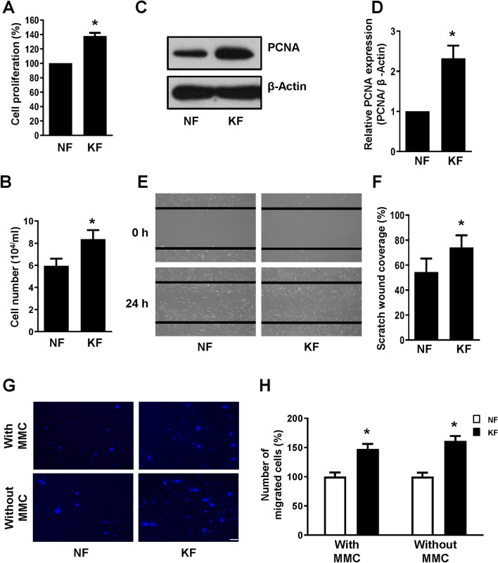
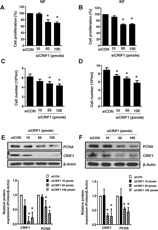
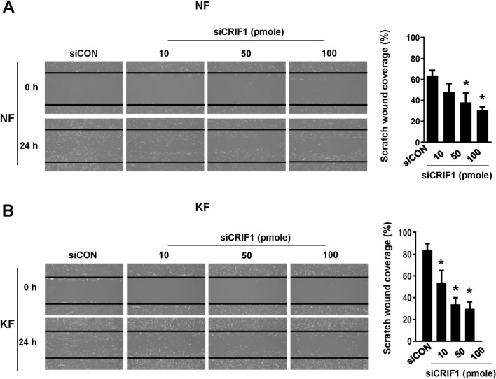
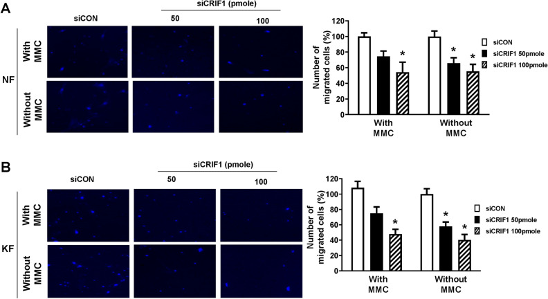
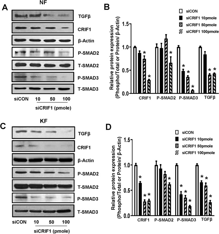
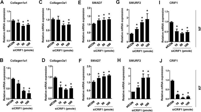
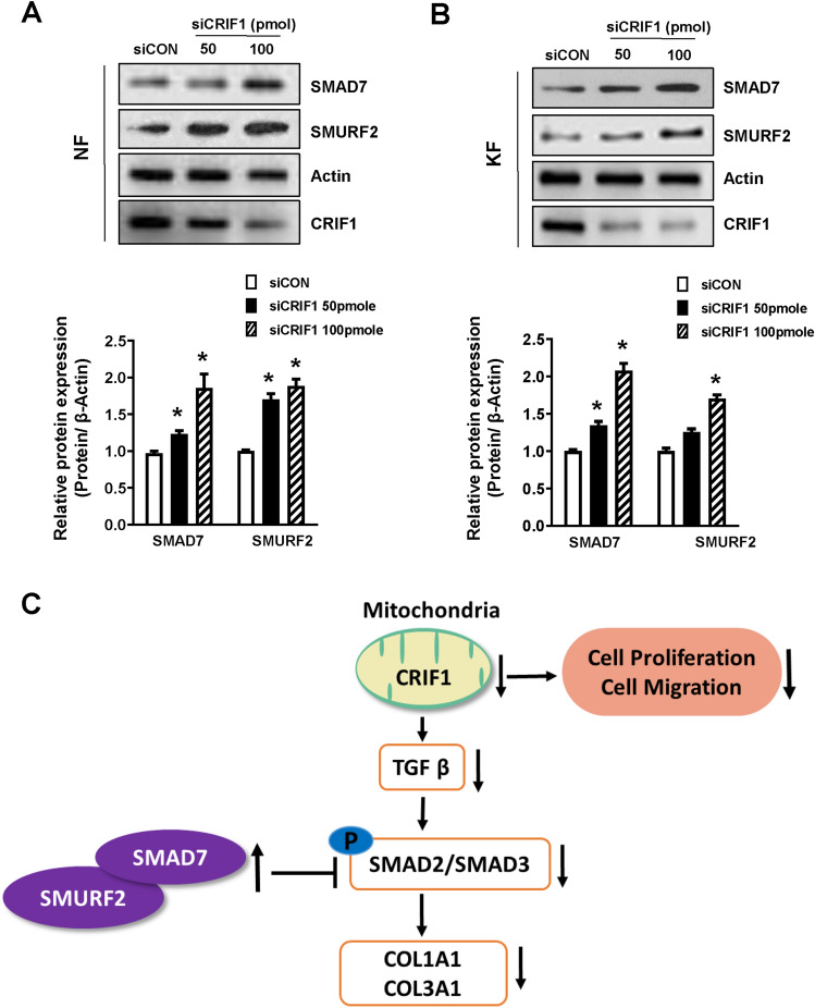
Keloids are a type of aberrant skin scarring characterized by excessive accumulation of collagen and extracellular matrix (ECM), arising from uncontrolled wound healing responses. While typically non-pathogenic, keloids are occasionally regarded as a form of benign tumor. CR6-interacting factor 1 (CRIF1) is a well-known CR6/GADD45-interacting protein, that has both nuclear and mitochondrial functions, and also exerts regulatory effects on cell growth and apoptosis. In this study, cell proliferation, cell migration, collagen production and TGF-β signaling was compared between normal fibroblasts (NFs) and keloid fibroblasts (KFs). Subsequently, the effects of CRIF1 deficiency were investigated in both NFs and KFs. Cell proliferation, cell migration, collagen production and protein expressions of TGF-β, phosphorylation of Smad2 and Smad3 were all found to be higher in KFs compared to NFs. CRIF1 deficiency in NFs and KFs inhibited cell proliferation, migration, and collagen production. In addition, phosphorylation of Smad2 and Smad3, which are transcription factors of collagen, was decreased. In contrast, mRNA expression levels of Smad7 and SMURF2, two important inhibitory proteins of Smad2/3, were increased, suggesting that CRIF1 may regulate collagen production. CRIF1 deficiency decreases the proliferation and migration of KFs, thereby inhibiting their overgrowth via the transforming growth factor-β (TGF-β)/Smad pathway. CRIF1 may therefore represent a potential therapeutic target in keloid pathogenesis.


Conflict of interest statement

The authors declare no competing interests.

Figures

Figure 1
Figure 1
Comparison of cell proliferation and cell migration between normal fibroblasts (NFs) and keloid fibroblasts (KFs). (A) NFs and KFs were cultured in 6-well plates for 24 h. After incubation, cells were treated with Cell Counting Kit-8 (CCK-8) solution, with cell proliferation measured based on absorbance at 450 nm. (B) NFs and KFs were cultured in 6-well plates for 24 h. After incubation, cells were harvested and ADAM-MC kit was used for cell counting. (C) Proliferating cell nuclear antigen (PCNA) protein expression, used as a marker of cell proliferation, was measured by western blot; β-actin was used as the loading control. Full-length blots are presented in Supplementary Fig. S1. (D) PCNA density was calculated using ImageJ software. (E) NFs and KFs were cultured in 6-well plates for 24 h followed by wounding for 24 h. Images were taken using a light microscope. (F) Quantification of wound closure was performed using ImageJ software. (G) Transwell assay was conducted to determine cell migration with or without MMC treatment. Scale bar 200 μm. (H) Quantification of the number of migrated cells was performed using ImageJ software. All data are presented as the mean ± SD of three independent experiments. *P < 0.05 relative to NFs.
Figure 2
Figure 2
Comparison of transforming growth factor-β (TGF-β)/Smad expression levels between normal fibroblasts (NFs) and keloid fibroblasts (KFs). (A) NFs and KFs were cultured in 6-well plates for 24 h. After 24 h incubation, cells were harvested, lysed, and analyzed by western blot to assess differences in protein levels of various proteins of the TGF-β/Smad signaling pathway and CRIF1; β-actin was used as the loading control. Full-length blots are presented in Supplementary Fig. S2. (B) Protein density was calculated using ImageJ software. The mRNA levels of (C) collagen 1A1 (COL1A1), (D) collagen 3A1 (COL3A1) (E) Smad7 and (F) SMURF2 were determined by quantitative polymerase chain reaction (qPCR) in NFs and KFs. All data are presented as the mean ± SD of three independent experiments. *P < 0.05 relative to NFs.
Figure 3
Figure 3
CR6-interacting factor 1 (CRIF1) downregulation leads to decreased cell proliferation in normal fibroblasts (NFs) and keloid fibroblasts (KFs). NFs were transfected with CRIF1 small interfering RNA (siRNA) in a dose-dependent manner for 48 h. (A) After incubation, cells were treated with CCK-8 solution, with cell proliferation measured based on absorbance at 450 nm. (C) After incubation, cells were harvested and ADAM-MC kit was used for cell counting. (E) PCNA protein levels were measured by Western blot; β-actin was used as the loading control. PCNA density was calculated using ImageJ software. Full-length blots are presented in Supplementary Fig. S3. KFs were transfected with CRIF1 small interfering RNA (siRNA) in a dose-dependent manner for 48 h. (B) After incubation, cells were treated with CCK-8 solution, with cell proliferation measured based on absorbance at 450 nm. (D) After incubation, cells were harvested and ADAM-MC kit was used for cell counting. (F) PCNA protein levels were measured by western blot; β-actin was used as the loading control. PCNA density was calculated using ImageJ software. Full-length blots are presented in Supplementary Fig. S4. All data are presented as means ± SD of three independent experiments. *P < 0.05 compared to siCON.
Figure 4
Figure 4
CR6-interacting factor 1 (CRIF1) downregulation leads to decreased cell migration measured by scratch wound healing assay in normal fibroblasts (NFs) and keloid fibroblasts (KFs). (A) NFs were transfected with CRIF1 siRNA in a dose-dependent manner for 24 h, followed by wounding for 24 h. (B) KFs were transfected with CRIF1 siRNA in a dose-dependent manner for 24 h, followed by wounding for 24 h. Images were taken using a light microscope. Quantification of wound closure was performed using ImageJ software. All data are presented as means ± SD of three independent experiments. *P < 0.05 compared to siCON.
Figure 5
Figure 5
CR6-interacting factor 1 (CRIF1) downregulation leads to decreased cell migration measured by transwell assay in normal fibroblasts (NFs) and keloid fibroblasts (KFs). (A) NFs were transfected with CRIF1 siRNA in a dose-dependent manner and transwell assay was conducted to determine cell migration with or without MMC treatment. Scale bar 200 μm. (B) KFs were transfected with CRIF1 siRNA in a dose-dependent manner and transwell assay was conducted to determine cell migration with or without MMC treatment. Scale bar 200 μm. Quantification of migrated cells was performed using ImageJ software. All data are presented as means ± SD of three independent experiments. *P < 0.05 compared to siCON.
Figure 6
Figure 6
The TGF-β/Smad pathway is altered by CRIF1 downregulation in normal fibroblasts (NFs) and keloid fibroblasts (KFs). (A) NFs were transfected with CRIF1 siRNA in a dose-dependent manner for 48 h. After 48 h incubation, cells were harvested, lysed, and analyzed by western blot to assess differences in protein levels of TGF-β, P-Smad2, and P-Smad3; β-actin was used as the loading control. Full-length blots are presented in Supplementary Fig. S5a. (B) Protein density was calculated using ImageJ software. (C) KFs were transfected with CRIF1 siRNA in a dose-dependent manner for 48 h. After 48 h incubation, cells were harvested, lysed, and analyzed by western blot to assess differences in protein levels of TGF-β, P-Smad2, and P-Smad3; β-actin was used as the loading control. Full-length blots are presented in Supplementary Fig. S5b. (D) Protein density was calculated using ImageJ software. All data are presented as the mean ± SD of three independent experiments. *P < 0.05 compared to siCON.
Figure 7
Figure 7
Expression of ECM components, Smad7 and SMURF2 mRNA levels are altered by CRIF1 downregulation in normal fibroblasts (NFs) and keloid fibroblasts (KFs). NFs were transfected with CRIF1 siRNA in a dose-dependent manner for 48 h. After 48 h incubation, mRNA expression levels of (A) COL1A1, (C) COL3A1 and (E) SMAD7, (G) SMURF2 and (I) CRIF1 were determined by qPCR. KFs were transfected with CRIF1 siRNA in a dose-dependent manner for 48 h. After 48 h incubation, mRNA expression levels of (B) COL1A1, (D) COL3A1 and (F) SMAD7, (H) SMURF2 and (J) CRIF1 were determined by qPCR. All data are presented as the mean ± SD of three independent experiments. *P < 0.05 compared to siCON.
Figure 8
Figure 8
Smad7 and SMURF2 protein levels are altered by CRIF1 downregulation in normal fibroblasts (NFs) and keloid fibroblasts (KFs). (A) NFs were transfected with CRIF1 siRNA in a dose-dependent manner for 48 h. After 48 h incubation, cells were harvested, lysed, and analyzed by western blot to assess differences in protein levels of SMAD7 and SMURF2; β-actin was used as the loading control. Protein density was calculated using ImageJ software. Full-length blots are presented in Supplementary Fig. S6a. (B) KFs were transfected with CRIF1 siRNA in a dose-dependent manner for 48 h. After 48 h incubation, cells were harvested, lysed, and analyzed by western blot to assess differences in protein levels of SMAD7 and SMURF2; β-actin was used as the loading control. Protein density was calculated using ImageJ software. Full-length blots are presented in Supplementary Fig. S6b. All data are presented as the mean ± SD of three independent experiments. *P < 0.05 compared to siCON. (C) Schematic representation of CRIF1 downregulation induced changes in the TGF-β/Smad signaling pathway leading to the inhibition of invasive KFs.

Similar articles

Cited by

References

    1. Li S, Liu W, Lei Y, Long J. Regulatory effects of electronic beam irradiation on mir-21/smad7-mediated collagen I synthesis in keloid-derived fibroblasts. Biol. Open. 2016;5:1567–1574. doi: 10.1242/bio.018770. - DOI - PMC - PubMed
    1. Dong X, Zhang C, Ma S, Wen H. Mast cell chymase in keloid induces profibrotic response via transforming growth factor-beta1/Smad activation in keloid fibroblasts. Int. J. Clin. Exp. Pathol. 2014;7:3596–3607. - PMC - PubMed
    1. Butler PD, Longaker MT, Yang GP. Current progress in keloid research and treatment. J. Am. Coll. Surg. 2008;206:731–741. doi: 10.1016/j.jamcollsurg.2007.12.001. - DOI - PubMed
    1. Seifert O, Mrowietz U. Keloid scarring: Bench and bedside. Arch. Dermatol. Res. 2009;301:259–272. doi: 10.1007/s00403-009-0952-8. - DOI - PubMed
    1. Shih B, Garside E, McGrouther DA, Bayat A. Molecular dissection of abnormal wound healing processes resulting in keloid disease. Wound Repair Regen. 2010;18:139–153. doi: 10.1111/j.1524-475X.2009.00553.x. - DOI - PubMed

Publication types

MeSH terms

LinkOut - more resources