. 2021 Jan 12;11(1):490.
doi: 10.1038/s41598-020-79434-4.

Human ex vivo lung perfusion: a novel model to study human lung diseases

Affiliations

Human ex vivo lung perfusion: a novel model to study human lung diseases

Nayra Cárdenes et al. Sci Rep. .

Abstract

Experimental animal models to predict physiological responses to injury and stress in humans have inherent limitations. Therefore, the development of preclinical human models is of paramount importance. Ex vivo lung perfusion (EVLP) has typically been used to recondition donor lungs before transplantation. However, this technique has recently advanced into a model to emulate lung mechanics and physiology during injury. In the present study, we propose that the EVLP of diseased human lungs is a well-suited preclinical model for translational research on chronic lung diseases. Throughout this paper, we demonstrate this technique's feasibility in pulmonary arterial hypertension (PAH), idiopathic pulmonary fibrosis (IPF), emphysema, and non-disease donor lungs not suitable for transplantation. In this study, we aimed to perfuse the lungs for 6 h with the EVLP system. This facilitated a robust and continuous assessment of airway mechanics, pulmonary hemodynamics, gas exchange, and biochemical parameters. We then collected at different time points tissue biopsies of lung parenchyma to isolate RNA and DNA to identify each disease's unique gene expression. Thus, demonstrating that EVLP could successfully serve as a clinically relevant experimental model to derive essential insights into pulmonary pathophysiology and various human lung diseases.


Conflict of interest statement

The authors declare no competing interests.

Figures

Figure 1
Figure 1
Human ex vivo lung perfusion. (A) Schematic of the experimental setup (previously published). (B) Kinetic analysis of Histopathology from Normal, PAH, Emphysema, and IPF Lungs during Ex Vivo Lung Perfusion (EVLP). Representative hematoxylin and eosin staining (20x) high-power magnification of lung parenchyma biopsy specimens at the beginning (0 h) and end of perfusion (6 h) shows maintenance of lung architecture. Quantification of the blind scoring of histology images is shown in bar graphs for time 0 and as fold change (FC) of end of EVLP over start. (C) Pulmonary artery pressure, PVR, peak Airway pressure, static lung compliance, and Peak Inspiratory Pressure throughout EVLP. (D) Gas exchange parameters including pO2, pCO2, HCO3, and pH and biochemical assessment of lung metabolism by measurement of glucose throughout perfusion in normal (red), emphysema (white), IPF (grey), and PAH (black) lungs at various timepoints on EVLP. *p < 0.05; **p < 0.01.
Figure 2
Figure 2
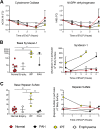
Soluble markers of cellular damage. (A) Mitochondrial DNA measured by qPCR of Cytochrome Oxidase and NADPH dehydrogenase, normalized to genomic DNA. The concentration of Syndecans (B) and Heparan sulfate (C) mRNA analysis of perfusate fluid measured by ELISA shown basal levels before EVLP (left) and through the course of the EVLP (right). *p < 0.05; **p < 0.01.
Figure 3
Figure 3
Cytokine measurements performed by multiplex ELISA are displayed for IL-6, IL-1β, IL-10, IFNγ, GM-CSF, IL-13, IL-7, and IL-8 (A-H). *p < 0.05; **p < 0.01; ****p < 0.0001.
Figure 4
Figure 4
Differential Gene expression (DEG) in lung tissue biopsies taken from PAH, IPF, Emphysema, and Normal lungs at time 0 h vs. 6 h during EVLP expressed as Venn diagrams. (A) Shows a Venn diagram with proportional representation of the number of genes, and (B) shows the number of DEG in each of the intersections.
Figure 5
Figure 5
Heatmap and volcano plot of differentially expressed genes before and after EVLP of all lung samples. FDR adjusted p value < 0.05.
Figure 6
Figure 6
Heatmap and volcano plots of differentially expressed genes before (green) and after EVLP (orange) in normal (A), IPF (B), PAH (C), and Emphysema (D). Raw p value < 0.01 and |logFC|> 1.
Figure 7
Figure 7
Heatmap and volcano plots of Differentially Expressed Genes before and after EVLP in IPF (A), Emphysema (B), and PAH (C) (green) compared to Normal (orange). Raw p value < 0.01 and |logFC|> 1.
Figure 8
Figure 8
Heat maps for differentially expressed genes (DEG) in Inflammatory Response, Hypoxia, Apoptosis, and UPR pathways. Top differentially expressed genes in the Inflammatory Response, Hypoxia, Apoptosis, and UPR pathways at baseline (yellow) and final state (grey) of EVLP for all group samples (Normal, Emphysema, IPF, and PAH). (A, C) FDR adjusted p value < 0.05. (B) raw p value < 0.01 and |logFC|> 1.

Similar articles

Cited by

References

    1. Soriano JB, Rodriguez-Roisin R. Chronic obstructive pulmonary disease overview: epidemiology, risk factors, and clinical presentation. Proc. Am. Thorac. Soc. 2011;8:363–367. doi: 10.1513/pats.201102-017RM. - DOI - PubMed
    1. Liu, T., De Los Santos, F. G. & Phan, S. H. The bleomycin model of pulmonary fibrosis. Methods Mol. Biol.1627, 27–42. 10.1007/978-1-4939-7113-8_2 (2017). - PubMed
    1. Moeller A, Ask K, Warburton D, Gauldie J, Kolb M. The bleomycin animal model: a useful tool to investigate treatment options for idiopathic pulmonary fibrosis? Int. J. Biochem. Cell Biol. 2008;40:362–382. doi: 10.1016/j.biocel.2007.08.011. - DOI - PMC - PubMed
    1. Herr C, et al. Combined exposure to bacteria and cigarette smoke resembles characteristic phenotypes of human COPD in a murine disease model. Exp. Toxicol. Pathol. 2015;67:261–269. doi: 10.1016/j.etp.2015.01.002. - DOI - PubMed
    1. Eltom, S., Stevenson, C. & Birrell, M. A. Cigarette smoke exposure as a model of inflammation associated with COPD. Curr. Protoc. Pharmacol.C10.1002/0471141755.ph0564s60 (2013). - PubMed

Publication types